

Wnt and Notch pathways have interrelated opposing roles on prostate progenitor cell proliferation and differentiation
- PMID: 21308863
- PMCID: PMC3148789
- DOI: 10.1002/stem.606
Wnt and Notch pathways have interrelated opposing roles on prostate progenitor cell proliferation and differentiation
Abstract
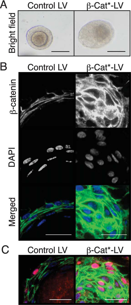
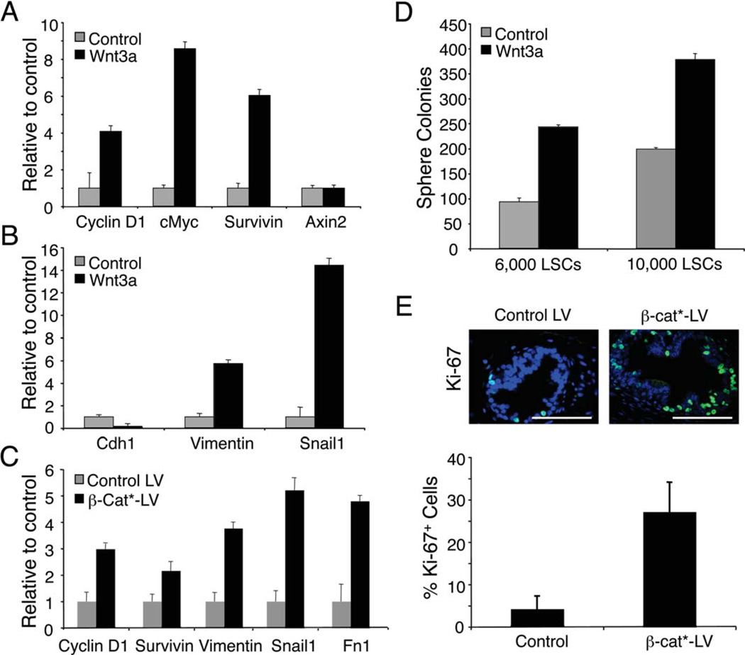
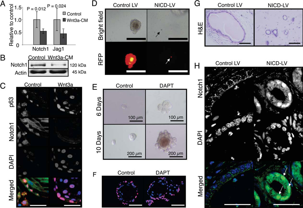
Tissue stem cells are capable of both self-renewal and differentiation to maintain a constant stem cell population and give rise to the plurality of cells within a tissue. Wnt signaling has been previously identified as a key mediator for the maintenance of tissue stem cells; however, possible cross-regulation with other developmentally critical signaling pathways involved in adult tissue homeostasis, such as Notch, is not well understood. By using an in vitro prostate stem cell colony ("prostasphere") formation assay and in vivo prostate reconstitution experiments, we demonstrate that Wnt pathway induction on Sca-1(+) CD49f(+) basal/stem cells (B/SCs) promotes expansion of the basal epithelial compartment with noticeable increases in "triple positive" (cytokeratin [CK] 5(+), CK8(+), p63(+)) prostate progenitor cells, concomitant with upregulation of known Wnt target genes involved in cell-cycle induction. Moreover, Wnt induction affects expression of epithelial-to-mesenchymal transition signature genes, suggesting a possible mechanism for priming B/SC to act as potential tumor-initiating cells. Interestingly, induction of Wnt signaling in B/SCs results in downregulation of Notch1 transcripts, consistent with its postulated antiproliferative role in prostate cells. In contrast, induction of Notch signaling in prostate progenitors inhibits their proliferation and disrupts prostasphere formation. In vivo prostate reconstitution assays further demonstrate that induction of Notch in B/SCs disrupts proper acini formation in cells expressing the activated Notch1 allele, Notch-1 intracellular domain. These data emphasize the importance of Wnt/Notch cross-regulation in adult stem cell biology and suggest that Wnt signaling controls the proliferation and/or maintenance of epithelial progenitors via modulation of Notch signaling.
Copyright © 2011 AlphaMed Press.
Conflict of interest statement
The authors indicate no potential conflicts of interest.
Figures


Similar articles
-
Parity induces differentiation and reduces Wnt/Notch signaling ratio and proliferation potential of basal stem/progenitor cells isolated from mouse mammary epithelium.Breast Cancer Res. 2013 Apr 29;15(2):R36. doi: 10.1186/bcr3419. Breast Cancer Res. 2013. PMID: 23621987 Free PMC article.
-
Tissue-dependent consequences of Apc inactivation on proliferation and differentiation of ciliated cell progenitors via Wnt and notch signaling.PLoS One. 2013 Apr 30;8(4):e62215. doi: 10.1371/journal.pone.0062215. Print 2013. PLoS One. 2013. PMID: 23646120 Free PMC article.
-
Tripartite interactions between Wnt signaling, Notch and Myb for stem/progenitor cell functions during intestinal tumorigenesis.Stem Cell Res. 2014 Nov;13(3 Pt A):355-66. doi: 10.1016/j.scr.2014.08.002. Epub 2014 Sep 28. Stem Cell Res. 2014. PMID: 25290188
-
Crosstalk between Wnt and Notch signaling in intestinal epithelial cell fate decision.J Gastroenterol. 2007 Sep;42(9):705-10. doi: 10.1007/s00535-007-2087-z. Epub 2007 Sep 25. J Gastroenterol. 2007. PMID: 17876539 Review.
-
Mitochondrial dynamics coordinate cell differentiation.Biochem Biophys Res Commun. 2018 May 27;500(1):59-64. doi: 10.1016/j.bbrc.2017.06.094. Epub 2017 Jun 17. Biochem Biophys Res Commun. 2018. PMID: 28634072 Review.
Cited by
-
Lgr4 is a key regulator of prostate development and prostate stem cell differentiation.Stem Cells. 2013 Nov;31(11):2492-505. doi: 10.1002/stem.1484. Stem Cells. 2013. PMID: 23897697 Free PMC article.
-
Notch signaling in prostate cancer: refining a therapeutic opportunity.Histol Histopathol. 2016 Feb;31(2):149-57. doi: 10.14670/HH-11-685. Epub 2015 Nov 2. Histol Histopathol. 2016. PMID: 26521657 Free PMC article. Review.
-
Notch and TGFβ form a reciprocal positive regulatory loop that suppresses murine prostate basal stem/progenitor cell activity.Cell Stem Cell. 2012 Nov 2;11(5):676-88. doi: 10.1016/j.stem.2012.07.003. Cell Stem Cell. 2012. PMID: 23122291 Free PMC article.
-
Chromatin effector Pygo2 mediates Wnt-notch crosstalk to suppress luminal/alveolar potential of mammary stem and basal cells.Cell Stem Cell. 2013 Jul 3;13(1):48-61. doi: 10.1016/j.stem.2013.04.012. Epub 2013 May 16. Cell Stem Cell. 2013. PMID: 23684539 Free PMC article.
-
Genome-wide tracts of homozygosity and exome analyses reveal repetitive elements with Barrets esophagus/esophageal adenocarcinoma risk.BMC Bioinformatics. 2019 Mar 14;20(Suppl 2):98. doi: 10.1186/s12859-019-2622-y. BMC Bioinformatics. 2019. PMID: 30871476 Free PMC article.
References
-
- Jemal A, Siegel R, Xu J, et al. Cancer statistics, 2010. CA Cancer J Clin. 2010;60:277–300. - PubMed
-
- Chesire DR, Ewing CM, Sauvageot J, et al. Detection and analysis of beta-catenin mutations in prostate cancer. Prostate. 2000;45:323–334. - PubMed
-
- Chesire DR, Isaacs WB. Beta-catenin signaling in prostate cancer: An early perspective. Endocr Relat Cancer. 2003;10:537–560. - PubMed
Publication types
MeSH terms
Substances
Grants and funding
LinkOut - more resources
Full Text Sources
Other Literature Sources
Medical
Research Materials
