

A semi-degradable composite scaffold for articular cartilage defects
- PMID: 21308980
- PMCID: PMC3139701
- DOI: 10.1002/jbm.a.33005
A semi-degradable composite scaffold for articular cartilage defects
Abstract
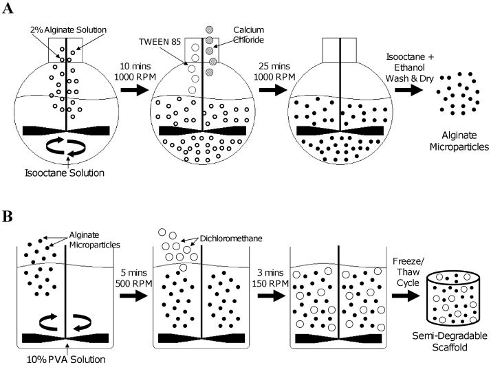
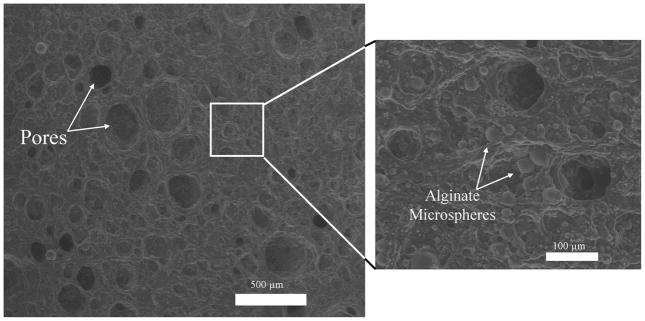
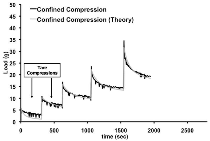
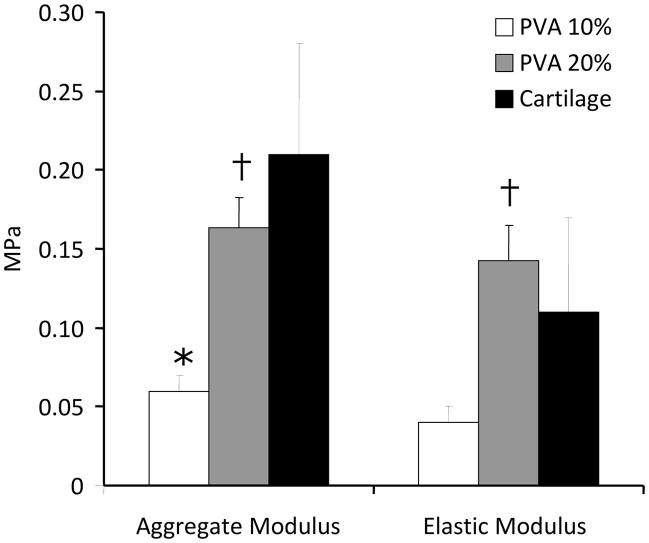
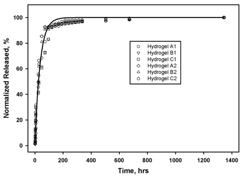
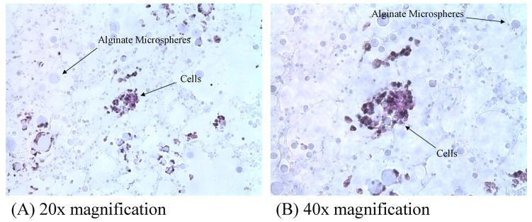
Few options exist to replace or repair damaged articular cartilage. The optimal solution that has been suggested is a scaffold that can carry load and integrate with surrounding tissues; but such a construct has thus far been elusive. The objectives of this study were to manufacture and characterize a nondegradable hydrated scaffold. Our hypothesis was that the polymer content of the scaffold can be used to control its mechanical properties, while an internal porous network augmented with biological agents can facilitate integration with the host tissue. Using a two-step water-in-oil emulsion process a porous polyvinyl alcohol (PVA) hydrogel scaffold combined with alginate microspheres was manufactured. The scaffold had a porosity of 11-30% with pore diameters of 107-187 μm, which readily allowed for movement of cells through the scaffold. Alginate microparticles were evenly distributed through the scaffold and allowed for the slow release of biological factors. The elastic modulus (Es ) and Poisson's ratio (υ), Aggregate modulus (Ha ) and dynamic modulus (ED ) of the scaffold were significantly affected by % PVA, as it varied from 10 to 20% wt/vol. Es and υ were similar to that of articular cartilage for both polymer concentrations, while Ha and ED were similar to that of cartilage only at 20% PVA. The ability to control scaffold mechanical properties, while facilitating cellular migration suggest that this scaffold is a potentially viable candidate for the functional replacement of cartilage defects.
Keywords: cartilage; osteoarthritis; polymer; scaffold; tissue engineering.
Copyright © 2011 Wiley Periodicals, Inc.
Figures
Similar articles
-
Characterization of a macroporous polyvinyl alcohol scaffold for the repair of focal articular cartilage defects.J Tissue Eng Regen Med. 2014 Feb;8(2):164-8. doi: 10.1002/term.1510. Epub 2012 May 2. J Tissue Eng Regen Med. 2014. PMID: 22549901 Free PMC article.
-
Poly (L-lactic acid) porous scaffold-supported alginate hydrogel with improved mechanical properties and biocompatibility.Int J Artif Organs. 2016 Oct 10;39(8):435-443. doi: 10.5301/ijao.5000516. Epub 2016 Sep 3. Int J Artif Organs. 2016. PMID: 27646631
-
Development of a novel alginate-polyvinyl alcohol-hydroxyapatite hydrogel for 3D bioprinting bone tissue engineered scaffolds.J Biomed Mater Res A. 2017 May;105(5):1457-1468. doi: 10.1002/jbm.a.36036. Epub 2017 Feb 25. J Biomed Mater Res A. 2017. PMID: 28187519
-
A novel method for the direct fabrication of growth factor-loaded microspheres within porous nondegradable hydrogels: controlled release for cartilage tissue engineering.J Control Release. 2012 Jan 10;157(1):39-45. doi: 10.1016/j.jconrel.2011.09.057. Epub 2011 Sep 10. J Control Release. 2012. PMID: 21930167
-
Semi-degradable porous poly (vinyl alcohol) hydrogel scaffold for cartilage repair: Evaluation of the initial and cell-cultured tribological properties.J Mech Behav Biomed Mater. 2017 Apr;68:163-172. doi: 10.1016/j.jmbbm.2017.02.001. Epub 2017 Feb 2. J Mech Behav Biomed Mater. 2017. PMID: 28183011
Cited by
-
Application of Alginate Hydrogels for Next-Generation Articular Cartilage Regeneration.Int J Mol Sci. 2022 Jan 20;23(3):1147. doi: 10.3390/ijms23031147. Int J Mol Sci. 2022. PMID: 35163071 Free PMC article. Review.
-
Tunable Alginate-Polyvinyl Alcohol Bioinks for 3D Printing in Cartilage Tissue Engineering.Gels. 2024 Dec 14;10(12):829. doi: 10.3390/gels10120829. Gels. 2024. PMID: 39727587 Free PMC article.
-
Porosity and cell preseeding influence electrospun scaffold maturation and meniscus integration in vitro.Tissue Eng Part A. 2013 Feb;19(3-4):538-47. doi: 10.1089/ten.TEA.2012.0052. Epub 2012 Nov 30. Tissue Eng Part A. 2013. PMID: 22994398 Free PMC article.
-
The Use of Nanomaterials in Tissue Engineering for Cartilage Regeneration; Current Approaches and Future Perspectives.Int J Mol Sci. 2020 Jan 14;21(2):536. doi: 10.3390/ijms21020536. Int J Mol Sci. 2020. PMID: 31947685 Free PMC article. Review.
-
Innovative hydrogel solutions for articular cartilage regeneration: a comprehensive review.Int J Surg. 2024 Dec 1;110(12):7984-8001. doi: 10.1097/JS9.0000000000002076. Int J Surg. 2024. PMID: 39236090 Free PMC article. Review.
References
-
- Mow VC, Ateshian GA, Spilker RL. Biomechanics of diarthrodial joints: a review of twenty years of progress. Journal of Biomechanical Engineering. 1993;115(4B):460. - PubMed
-
- Gilley JS, Gelman MI, Edson DM, Metcalf RW. Chondral fractures of the knee. Arthrographic, arthroscopic, and clinical manifestations. Radiology. 1981;138(1):51. - PubMed
-
- Shelbourne KD, Jari S, Gray T. Outcome of untreated traumatic articular cartilage defects of the knee: a natural history study. The Journal of bone and joint surgery. American volume. 2003;85-A(Suppl 2):8. - PubMed
-
- Buckwalter JA, Mankin HJ. Articular cartilage: tissue design and chondrocyte-matrix interactions. Instructional course lectures. 1998;47:477. - PubMed
-
- Mankin HJ, Mow VC, Buckwalter JA, Iannotti JP, Ratcliffe A. Form and Function of Articular Cartilage. In: Simon SR, editor. Orthopaedic Basic Science. Rosemont, Ill: American Academy of Orthopaedic Surgeons; 1994. pp. 1–44. - PubMed
Publication types
MeSH terms
Substances
Grants and funding
LinkOut - more resources
Full Text Sources
Other Literature Sources
Miscellaneous
