Case Reports
. 2011 Apr;32(4):445-55.
doi: 10.1002/humu.21462. Epub 2011 Mar 1.

New insights into the pathogenesis of autosomal-dominant cutis laxa with report of five ELN mutations

Affiliations
Case Reports

New insights into the pathogenesis of autosomal-dominant cutis laxa with report of five ELN mutations

Bert Callewaert et al. Hum Mutat. 2011 Apr.

Abstract

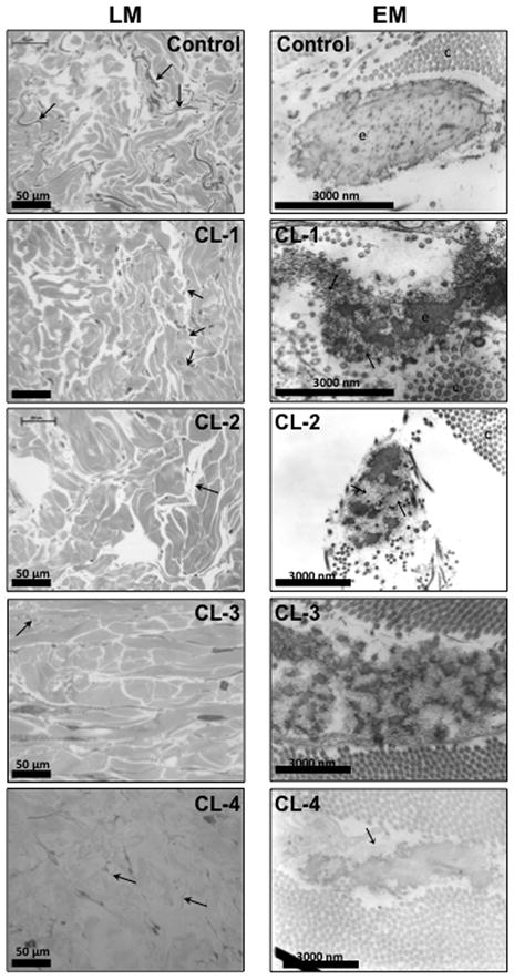
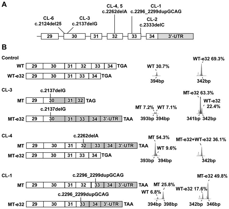
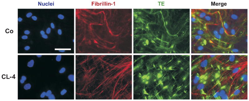
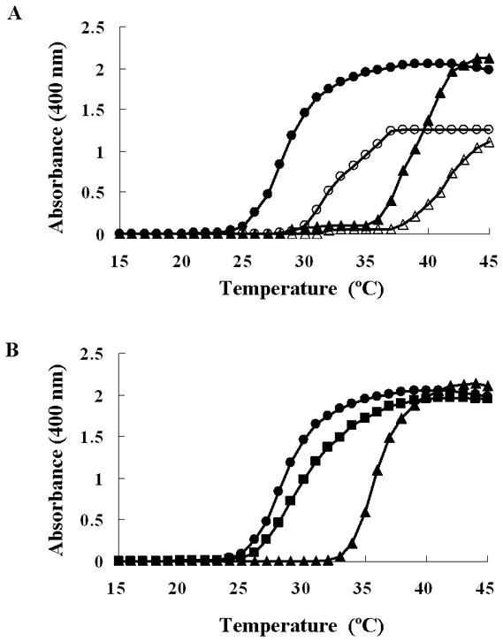
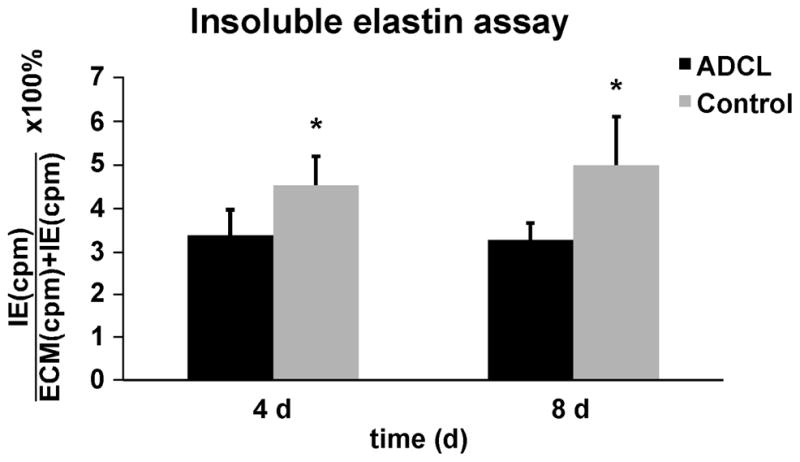
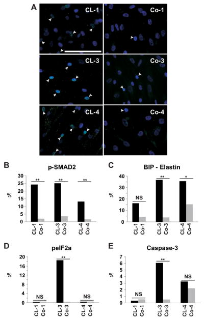
Autosomal dominant cutis laxa (ADCL) is characterized by a typical facial appearance and generalized loose skin folds, occasionally associated with aortic root dilatation and emphysema. We sequenced exons 28-34 of the ELN gene in five probands with ADCL features and found five de novo heterozygous mutations: c.2296_2299dupGCAG (CL-1), c.2333delC (CL-2), c.2137delG (CL-3), c.2262delA (monozygotic twin CL-4 and CL-5), and c.2124del25 (CL-6). Four probands (CL-1,-2,-3,-6) presented with progressive aortic root dilatation. CL-2 and CL-3 also had bicuspid aortic valves. CL-2 presented with severe emphysema. Electron microscopy revealed elastic fiber fragmentation and diminished dermal elastin deposition. RT-PCR studies showed stable mutant mRNA in all patients. Exon 32 skipping explains a milder phenotype in patients with exon 32 mutations. Mutant protein expression in fibroblast cultures impaired deposition of tropoelastin onto microfibril-containing fibers, and enhanced tropoelastin coacervation and globule formation leading to lower amounts of mature, insoluble elastin. Mutation-specific effects also included endoplasmic reticulum stress and increased apoptosis. Increased pSMAD2 staining in ADCL fibroblasts indicated enhanced transforming growth factor beta (TGF-β) signaling. We conclude that ADCL is a systemic disease with cardiovascular and pulmonary complications, associated with increased TGF-β signaling and mutation-specific differences in endoplasmic reticulum stress and apoptosis.


Figures

Figure 1
Figure 1
Clinical presentation of patients CL-1, CL-2, CL-3, CL-4 and 5, and CL-6. Note the variable severity of generalized cutis laxa, and the typical facial characteristics including a premature aged appearance and a long philtrum (CL-3, CL-4, CL-5) and large ears (CL-4, CL-5).
Figure 2
Figure 2
Light microscopic (LM) images of semi-thin methylene blue-stained sections of skin biopsies from a control (female, 15 years old), CL-1, CL-2, CL-3, and CL-4. Using this method, elastic fibers are stained darker than collagen bundles. Arrows indicate elastic fibers (dark grey). Elastic fibers are scarce in each ADCL sample. Electron microscopic (EM) images of a control sample, CL-1, CL-2, CL-3 and CL-4; c, collagen fibers; e, elastin; arrow, microfibrils not embedded in elastin. Patients show elastic fibers with diminished amounts and abnormal morphology of amorphous elastin, including extensive branching and fragmentation and a lack of proper association with the microfibrils. The electron density increases from inner to outer regions of the elastic material and there are separate elastic globules. In patient CL-4, similar, but milder abnormalities are present.
Figure 3
Figure 3
A: Schematic representation of the 3′-end of ELN with the location of the mutations identified in each patient. The coding exons are drawn to scale but the 3′-untranslated region (3′-UTR, hatched) and the introns are not. B: Schematic representation of the structure of wildtype (WT) and mutant (MT) mRNA products with semi-quantitative analysis. ELN transcripts in ADCL and control fibroblasts were studied using a oligonucleotide sense and antisense primer complementary to exon 29 and the 3′-UTR, respectively (Szabo, et al., 2006). M13 fluorescently labeled PCR-based amplicons were run on a genetic analyzer and data were processed using Genescan software (Applied Biosystems). Electropherograms are shown next to schematic representations of mRNA species identified in the control and in each patient. Cross-hatched bars indicate sequences coding for missense or read-through peptides generated by each frame shift mutation. Normal ELN sequence is shown by open bars. For patients only the products of the mutant allele are drawn, but electropherograms show both WT and MT products. Identities, sizes and % abundance relative to total ELN mRNA in each sample are shown next to each peak. In control fibroblasts exon 32 is spliced out (-e32) in about 75% of the transcripts (peak at 342 nucleotides). In CL-3 there is markedly higher levels of the mutant mRNA in transcripts lacking exon 32. Full-length transcripts of the wild type and mutant allele are present at low quantities. In patient CL-4, there is higher abundance of mutant mRNA in the full-length transcript (transcripts without exon 32 are all wild type). In patient CL-1, increased levels of the 4-nucleotide larger mutant mRNA is observed.
Figure 4
Figure 4
Representative colocalization immunofluorescent staining for fibrillin-1 (red) and TE (green) of non-permeabilized cell cultures of patient CL-4 and a matching control (Co). In the patient, elastin was less densely deposited in fibers, and showed significantly enhanced aggregation in clumps. Nuclei were stained blue. Scale bar: 25 μm.
Figure 5
Figure 5
A: Coacervation of TE and fmTE. TE (triangles) and fmTE (circles) were diluted to a concentration of 6.25 μM (open symbols) and 12.5 μM (closed symbols) in PBS. Light scattering was monitored every 0.5 min while raising the temperature form 15 °C to 45 °C. B: Coacervation of a mixture of TE and fmTE was studied at a concentration of 6.25 μM each in PBS. As reference, pure TE and fmTE protein solutions were evaluated at concentration of 12.5 μM in PBS.
Figure 6
Figure 6
Biochemically isolated insoluble elastin was measured form metabolically labeled cell cultures and normalized to all intra- and extracellular proteins in the cell layer (as a measure for cell metabolism) and cell count. Bars represent the averaged values of insoluble elastin in all ADCL and matched control fibroblasts. There was a significant reduction of mature, crosslinked, insoluble elastin after 4 and 8 days in ADCL patients. *p value < 0.01 using the paired one-tailed student T-test. Error bars show the standard deviation of the mean.
Figure 7
Figure 7
A: p-SMAD2 staining in fibroblast cultures. All patients (CL-1, CL-3, and CL-4) showed increased proportion of p-SMAD2 positive cells (p<0.001) indicating enhanced TGFβ signaling compared to matching controls (Co-1, Co-3 and Co-4). Scale bar: 50 μm. B: Quantitative morphometry of pSMAD2 staining in fibroblasts of ADCL patients and their matching controls. Quantitative morphometry of C: BiP; D: peIF2α; E: caspase-3 staining in fibroblasts of ADCL patients and matching controls. Patients CL-3 and CL-4, but not patient CL- 1, showed upregulation of the chaperone BiP co-localizing with TE in the ER. peIF2α and caspase-3 are significantly upregulated in patient CL-3, but not in CL-1 or CL-4. **: p<0.001; *: p<0.01; NS: not significant.

Similar articles

Cited by

References

    1. Basel-Vanagaite L, Sarig O, Hershkovitz D, Fuchs-Telem D, Rapaport D, Gat A, Isman G, Shirazi I, Shohat M, Enk CD, Birk E, Kohlhase J, Matysiak-Scholze U, Maya I, Knopf C, Peffekoven A, Hennies HC, Bergman R, Horowitz M, Ishida-Yamamoto A, Sprecher E. RIN2 deficiency results in macrocephaly, alopecia, cutis laxa, and scoliosis: MACS syndrome. Am J Hum Genet. 2009;85:254–263. - PMC - PubMed
    1. Bicknell LS, Pitt J, Aftimos S, Ramadas R, Maw MA, Robertson SP. A missense mutation in ALDH18A1, encoding Delta1-pyrroline-5-carboxylate synthase (P5CS), causes an autosomal recessive neurocutaneous syndrome. Eur J Hum Genet. 2008;16:1176–1186. - PubMed
    1. Broekelmann TJ, Kozel BA, Ishibashi H, Werneck CC, Keeley FW, Zhang L, Mecham RP. Tropoelastin interacts with cell-surface glycosaminoglycans via its COOH-terminal domain. J Biol Chem. 2005;280(49):40939–40947. - PubMed
    1. Brown FR, 3rd, Holbrook KA, Byers PH, Stewart D, Dean J, Pyeritz RE. Cutis laxa. Johns Hopkins Med J. 1982;150:148–153. - PubMed
    1. Brown-Augsburger P, Broekelmann T, Mecham L, Mercer R, Gibson MA, Cleary EG, Abrams WR, Rosenbloom J, Mecham RP. Microfibril-associated glycoprotein binds to the carboxyl-terminal domain of tropoelastin and is a substrate for transglutaminase. J Biol Chem. 1994;269:28443–28449. - PubMed

Publication types