

The eye as an organizer of craniofacial development
- PMID: 21309065
- PMCID: PMC3690320
- DOI: 10.1002/dvg.20716
The eye as an organizer of craniofacial development
Erratum in
- Genesis. 2013 Apr;51(4):293
Abstract
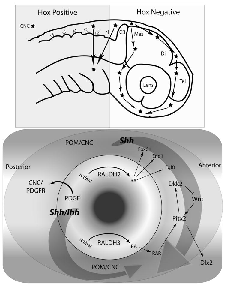
The formation and invagination of the optic stalk coincides with the migration of cranial neural crest (CNC) cells, and a growing body of data reveals that the optic stalk and CNC cells communicate to lay the foundations for periocular and craniofacial development. Following migration, the interaction between the developing eye and surrounding periocular mesenchyme (POM) continues, leading to induction of transcriptional regulatory cascades that regulate craniofacial morphogenesis. Studies in chick, mice, and zebrafish have revealed a remarkable level of genetic and mechanistic conservation, affirming the power of each animal model to shed light on the broader morphogenic process. This review will focus on the role of the developing eye in orchestrating craniofacial morphogenesis, utilizing morphogenic gradients, paracrine signaling, and transcriptional regulatory cascades to establish an evolutionarily-conserved facial architecture. We propose that in addition to the forebrain, the eye functions during early craniofacial morphogenesis as a key organizer of facial development, independent of its role in vision.
Copyright © 2011 Wiley-Liss, Inc.
Figures
Similar articles
-
A zebrafish model of axenfeld-rieger syndrome reveals that pitx2 regulation by retinoic acid is essential for ocular and craniofacial development.Invest Ophthalmol Vis Sci. 2012 Jan 3;53(1):7-22. doi: 10.1167/iovs.11-8494. Invest Ophthalmol Vis Sci. 2012. PMID: 22125274 Free PMC article.
-
Pax6 regulates craniofacial form through its control of an essential cephalic ectodermal patterning center.Genesis. 2011 Apr;49(4):307-25. doi: 10.1002/dvg.20724. Genesis. 2011. PMID: 21309073
-
Sox9 function in craniofacial development and disease.Genesis. 2011 Apr;49(4):200-8. doi: 10.1002/dvg.20717. Epub 2011 Apr 1. Genesis. 2011. PMID: 21309066 Free PMC article. Review.
-
Regulation of PP2A activity by Mid1 controls cranial neural crest speed and gangliogenesis.Mech Dev. 2012 Jan-Feb;128(11-12):560-76. doi: 10.1016/j.mod.2012.01.002. Epub 2012 Jan 20. Mech Dev. 2012. PMID: 22285438
-
Collective cell migration of the cephalic neural crest: the art of integrating information.Genesis. 2011 Apr;49(4):164-76. doi: 10.1002/dvg.20700. Epub 2011 Jan 24. Genesis. 2011. PMID: 21157935 Review.
Cited by
-
A zebrafish model of axenfeld-rieger syndrome reveals that pitx2 regulation by retinoic acid is essential for ocular and craniofacial development.Invest Ophthalmol Vis Sci. 2012 Jan 3;53(1):7-22. doi: 10.1167/iovs.11-8494. Invest Ophthalmol Vis Sci. 2012. PMID: 22125274 Free PMC article.
-
The human brain and face: mechanisms of cranial, neurological and facial development revealed through malformations of holoprosencephaly, cyclopia and aberrations in chromosome 18.J Anat. 2015 Sep;227(3):255-67. doi: 10.1111/joa.12343. J Anat. 2015. PMID: 26278930 Free PMC article.
-
Overview of Head Muscles with Special Emphasis on Extraocular Muscle Development.Adv Anat Embryol Cell Biol. 2023;236:57-80. doi: 10.1007/978-3-031-38215-4_3. Adv Anat Embryol Cell Biol. 2023. PMID: 37955771
-
Early lens ablation causes dramatic long-term effects on the shape of bones in the craniofacial skeleton of Astyanax mexicanus.PLoS One. 2012;7(11):e50308. doi: 10.1371/journal.pone.0050308. Epub 2012 Nov 30. PLoS One. 2012. PMID: 23226260 Free PMC article.
-
Neural crest derivatives in ocular development: discerning the eye of the storm.Birth Defects Res C Embryo Today. 2015 Jun;105(2):87-95. doi: 10.1002/bdrc.21095. Epub 2015 Jun 4. Birth Defects Res C Embryo Today. 2015. PMID: 26043871 Free PMC article. Review.
References
-
- Abe M, Maeda T, Wakisaka S. Retinoic acid affects craniofacial patterning by changing Fgf8 expression in the pharyngeal ectoderm. Dev Growth Differ. 2008;50:717–729. - PubMed
-
- Ahlgren SC, Bronner-Fraser M. Inhibition of sonic hedgehog signaling in vivo results in craniofacial neural crest cell death. Curr Biol. 1999;9:1304–1314. - PubMed
-
- Alunni A, Menuet A, Candal E, Penigault JB, Jeffery WR, Retaux S. Developmental mechanisms for retinal degeneration in the blind cavefish Astyanax mexicanus. J Comp Neurol. 2007;505:221–233. - PubMed
-
- Beck AE, Hudgins L, Hoyme HE. Autosomal dominant microtia and ocular coloboma: new syndrome or an extension of the oculo-auriculo-vertebral spectrum? Am J Med Genet A. 2005;134:359–362. - PubMed
Publication types
MeSH terms
Substances
Grants and funding
LinkOut - more resources
Full Text Sources
Medical
