Mechanisms involved in extraterritorial facial pain following cervical spinal nerve injury in rats
- PMID: 21310020
- PMCID: PMC3048571
- DOI: 10.1186/1744-8069-7-12
Mechanisms involved in extraterritorial facial pain following cervical spinal nerve injury in rats
Abstract
Background: The aim of this study is to clarify the neural mechanisms underlying orofacial pain abnormalities after cervical spinal nerve injury. Nocifensive behavior, phosphorylated extracellular signal-regulated kinase (pERK) expression and astroglial cell activation in the trigeminal spinal subnucleus caudalis (Vc) and upper cervical spinal dorsal horn (C1-C2) neurons were analyzed in rats with upper cervical spinal nerve transection (CNX).
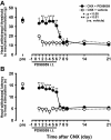
Results: The head withdrawal threshold to mechanical stimulation of the lateral facial skin and head withdrawal latency to heating of the lateral facial skin were significantly lower and shorter respectively in CNX rats compared to Sham rats. These nocifensive effects were apparent within 1 day after CNX and lasted for more than 21 days. The numbers of pERK-like immunoreactive (LI) cells in superficial laminae of Vc and C1-C2 were significantly larger in CNX rats compared to Sham rats following noxious and non-noxious mechanical or thermal stimulation of the lateral facial skin at day 7 after CNX. Two peaks of pERK-LI cells were observed in Vc and C1-C2 following mechanical and heat stimulation of the lateral face. The number of pERK-LI cells in C1-C2 was intensity-dependent and increased when the mechanical and heat stimulations of the face were increased. The decrements of head withdrawal latency to heat and head withdrawal threshold to mechanical stimulation were reversed during intrathecal (i.t.) administration of MAPK/ERK kinase 1/2 inhibitor PD98059. The area of activated astroglial cells was significantly higher in CNX rats (at day 7 after CNX). The heat and mechanical nocifensive behaviors were significantly depressed and the number of pERK-LI cells in Vc and C1-C2 following noxious and non-noxious mechanical stimulation of the face was also significantly decreased following i.t. administration of the astroglial inhibitor fluoroacetate.
Conclusions: The present findings have demonstrated that mechanical allodynia and thermal hyperalgesia occur in the lateral facial skin after CNX and also suggest that ERK phosphorylation of Vc and C1-C2 neurons and astroglial cell activation are involved in orofacial extraterritorial pain following cervical nerve injury.
Figures


Similar articles
-
Mechanisms involved in an increment of multimodal excitability of medullary and upper cervical dorsal horn neurons following cutaneous capsaicin treatment.Mol Pain. 2008 Nov 19;4:59. doi: 10.1186/1744-8069-4-59. Mol Pain. 2008. PMID: 19019214 Free PMC article.
-
Organization of hyperactive microglial cells in trigeminal spinal subnucleus caudalis and upper cervical spinal cord associated with orofacial neuropathic pain.Brain Res. 2012 Apr 27;1451:74-86. doi: 10.1016/j.brainres.2012.02.023. Epub 2012 Feb 17. Brain Res. 2012. PMID: 22459040
-
ERK-GluR1 phosphorylation in trigeminal spinal subnucleus caudalis neurons is involved in pain associated with dry tongue.Mol Pain. 2016 Apr 26;12:1744806916641680. doi: 10.1177/1744806916641680. Print 2016. Mol Pain. 2016. PMID: 27118769 Free PMC article.
-
Neuron-glia interaction is a key mechanism underlying persistent orofacial pain.J Oral Sci. 2017;59(2):173-175. doi: 10.2334/josnusd.16-0858. J Oral Sci. 2017. PMID: 28637974 Review.
-
Peripheral and Central Mechanisms of Persistent Orofacial Pain.Front Neurosci. 2019 Nov 13;13:1227. doi: 10.3389/fnins.2019.01227. eCollection 2019. Front Neurosci. 2019. PMID: 31798407 Free PMC article. Review.
Cited by
-
Generalized Extension of Referred Trigeminal Pain due to Greater Occipital Nerve Entrapment.Case Rep Neurol Med. 2023 Nov 11;2023:1099222. doi: 10.1155/2023/1099222. eCollection 2023. Case Rep Neurol Med. 2023. PMID: 38025301 Free PMC article.
-
Chronic Orofacial Pain: Models, Mechanisms, and Genetic and Related Environmental Influences.Int J Mol Sci. 2021 Jul 1;22(13):7112. doi: 10.3390/ijms22137112. Int J Mol Sci. 2021. PMID: 34281164 Free PMC article. Review.
-
The Role of Stress in Burning Mouth Syndrome Triggered by Dental Treatments: A Two-Step Hypothesis.J Oral Rehabil. 2025 Jul;52(7):1001-1014. doi: 10.1111/joor.13964. Epub 2025 Mar 26. J Oral Rehabil. 2025. PMID: 40143520 Free PMC article.
-
Metabotropic glutamate receptor 5 contributes to inflammatory tongue pain via extracellular signal-regulated kinase signaling in the trigeminal spinal subnucleus caudalis and upper cervical spinal cord.J Neuroinflammation. 2012 Nov 27;9:258. doi: 10.1186/1742-2094-9-258. J Neuroinflammation. 2012. PMID: 23181395 Free PMC article.
-
Involvement of peripheral ionotropic glutamate receptors in orofacial thermal hyperalgesia in rats.Mol Pain. 2011 Sep 28;7:75. doi: 10.1186/1744-8069-7-75. Mol Pain. 2011. PMID: 21952000 Free PMC article.
References
-
- Sessle BJ. Neural mechanisms and pathways in craniofacial pain. Can J Neurol Sci. 1999;26(Suppl 3):S7–11. - PubMed
-
- Hsieh GC, Pai M, Chandran P, Hooker BA, Zhu CZ, Salyers AK, Wensink EJ, Zhan C, Carroll WA, Dart MJ. et al.Central and peripheral sites of action for CB(2) receptor mediated analgesic activity in chronic inflammatory and neuropathic pain models in rats. Br J Pharmacol. 2011;162(2):428–40. doi: 10.1111/j.1476-5381.2010.01046.x. - DOI - PMC - PubMed
Publication types
MeSH terms
Substances
Grants and funding
LinkOut - more resources
Full Text Sources
Medical
Miscellaneous

