. 2011 Feb 10:11:65.
doi: 10.1186/1471-2407-11-65.

The level of claudin-7 is reduced as an early event in colorectal carcinogenesis

Affiliations

The level of claudin-7 is reduced as an early event in colorectal carcinogenesis

Jette Bornholdt et al. BMC Cancer. .

Abstract

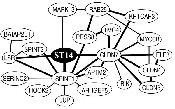
Background: Compromised epithelial barriers are found in dysplastic tissue of the gastrointestinal tract. Claudins are transmembrane proteins important for tight junctions. Claudins regulate the paracellular transport and are crucial for maintaining a functional epithelial barrier. Down-regulation of the oncogenic serine protease, matriptase, induces leakiness in epithelial barriers both in vivo and in vitro. We found in an in-silico search tight co-regulation between matriptase and claudin-7 expression. We have previously shown that the matriptase expression level decreases during colorectal carcinogenesis. In the present study we investigated whether claudin-7 expression is likewise decreased during colorectal carcinogenesis, thereby causing or contributing to the compromised epithelial leakiness of dysplastic tissue.

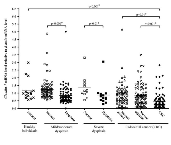
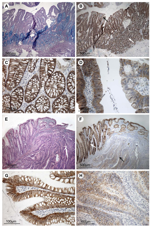
Methods: The mRNA level of claudin-7 (CLDN7) was determined in samples from 18 healthy individuals, 100 individuals with dysplasia and 121 colorectal cancer patients using quantitative real time RT-PCR. In addition, immunohistochemical stainings were performed on colorectal adenomas and carcinomas, to confirm the mRNA findings.

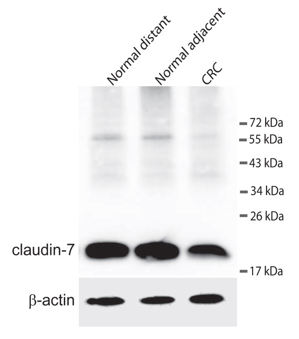
Results: A 2.7-fold reduction in the claudin-7 mRNA level was found when comparing the biopsies from healthy individuals with the biopsies of carcinomas (p < 0.001). Reductions in the claudin-7 mRNA levels were also detected in mild/moderate dysplasia (p < 0.001), severe dysplasia (p < 0.01) and carcinomas (p < 0.01), compared to a control sample from the same individual. The decrease at mRNA level was confirmed at the protein level by immunohistochemical stainings.

Conclusions: Our results show that the claudin-7 mRNA level is decreased already as an early event in colorectal carcinogenesis, probably contributing to the compromised epithelial barrier in adenomas.


Figures

Figure 1
Figure 1
In-silico analysis showing the ST14 co-expressed gene network. The figure shows a modified version of the analysis performed using the COXPRESdb version 4.0 http://coxpresdb.jp/. Each gene is represented by a node. Lines between nodes indicate co-expression. The genes with direct ST14 co-expression are marked with a line to the ST14 node. These genes are SPINT1 (encoding HAI-1), CLDN7 (encoding claudin-7), LSR (encoding lipolysis stimulated lipoprotein receptor) and MAPK13 (encoding mitogen-activated protein kinase 13). Bold lines indicate a mutual ratio (MR) below 5 and normal lines indicate a MR between 5 and 30.
Figure 2
Figure 2
Claudin-7 mRNA levels in healthy individuals, individuals with dysplasia and individuals with carcinomas as determined by real-time RT-PCR. Samples from healthy individuals (cross), normal (open circle) and affected tissue (filled circle) from individuals with mild/moderate dysplasia, normal (open square) and affected tissue (filled square) from individuals with severe dysplasia, normal adjacent (open triangle), normal distant (open triangle) and carcinomatous tissue (filled diamond) from colorectal cancer patients were analysed for claudin-7 mRNA levels relative to the β-actin mRNA levels. The horizontal line represents the mean values. The p-value indicated with a ¤ is calculated using Kruskal Wallis and Dunn's Multiple Comparison test and p-values marked with a * are calculated using Paired Student's t-test.
Figure 3
Figure 3
Western blot detecting claudin-7 and ß-actin. The upper panel shows claudin-7 in samples from a colorectal cancer patient. Normal distant is a sample taken as far away from the tumour as possible in the surgically removed tissue. Normal adjacent is a sample taken just adjacent to the tumour and CRC is from the cancer itself. The lower panel shows the loading control (ß-actin).
Figure 4
Figure 4
Immunohistochemical staining for claudin-7 in colorectal adenomas, carcinomas and adjacent normal tissue. The top four pictures are taken from an individual with dysplasia but no record of carcinoma and the bottom four pictures are from a patient with colorectal cancer. A) Tissue section including both normal mucosa and dysplastic tissue, stained with PAS/Alcian. B) Neighbouring tissue section stained for claudin-7. The white arrow indicates mucosa of normal appearance and the black arrow indicates dysplastic tissue. C) High magnification of the normal mucosa from B, showing staining mainly at the basolateral cell membranes of the epithelial cells. D) High magnification of the dysplastic tissue from B showing a patchy staining pattern with areas of low and normal staining. E) Tissue section with cancerous tissue to the right and normal mucosa to the left, stained with Hematoxylin and Eosin. F) Neighbouring tissue section stained for claudin-7. The white arrow indicates the mucosa with normal appearance. The black arrow points out carcinomatous tissue. G) High magnification of histologically normally appearing mucosa from F, showing staining mainly localised to the basolateral cell membranes of the surface epithelial cells. H) High magnification of carcinomatous tissue showing faint, patchy staining of the epithelial strands of tumour tissue. Scale bars: 500 μm (A + B + E + F), 100 μm (C + D + G + H).

References

    1. Soler AP, Miller RD, Laughlin KV, Carp NZ, Klurfeld DM, Mullin JM. Increased tight junctional permeability is associated with the development of colon cancer. Carcinogenesis. 1999;20:1425–1431. doi: 10.1093/carcin/20.8.1425. - DOI - PubMed
    1. Angelow S, Ahlstrom R, Yu AS. Biology of claudins. Am J Physiol Renal Physiol. 2008;295:F867–F876. doi: 10.1152/ajprenal.90264.2008. - DOI - PMC - PubMed
    1. Martin TA, Jiang WG. Loss of tight junction barrier function and its role in cancer metastasis. Biochim Biophys Acta. 2009;1788:872–891. doi: 10.1016/j.bbamem.2008.11.005. - DOI - PubMed
    1. Gonzalez-Mariscal L, Betanzos A, Nava P, Jaramillo BE. Tight junction proteins. Prog Biophys Mol Biol. 2003;81:1–44. doi: 10.1016/S0079-6107(02)00037-8. - DOI - PubMed
    1. Tsukita S, Furuse M, Itoh M. Multifunctional strands in tight junctions. Nat Rev Mol Cell Biol. 2001;2:285–293. doi: 10.1038/35067088. - DOI - PubMed

Publication types

MeSH terms