. 2011 Feb 11;88(2):216-25.
doi: 10.1016/j.ajhg.2011.01.004.

Identification and characterization of an inborn error of metabolism caused by dihydrofolate reductase deficiency

Affiliations

Identification and characterization of an inborn error of metabolism caused by dihydrofolate reductase deficiency

Siddharth Banka et al. Am J Hum Genet. .

Abstract

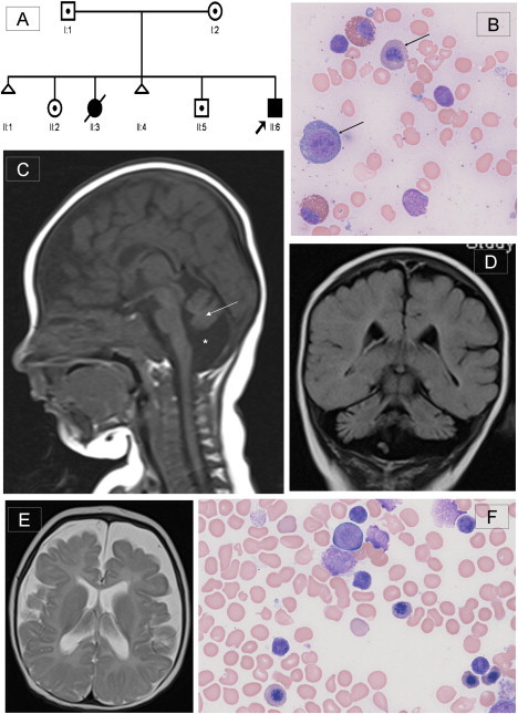
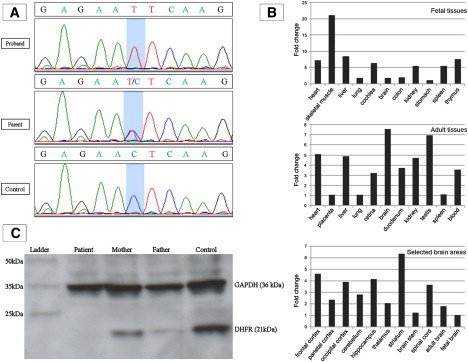
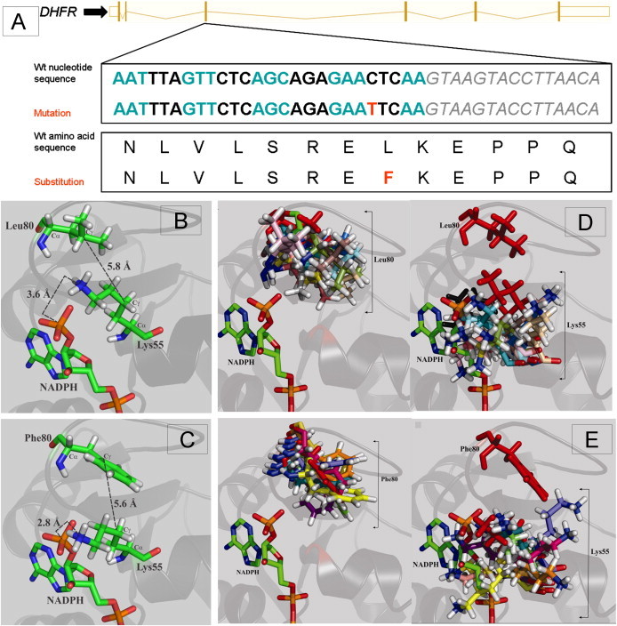
Dihydrofolate reductase (DHFR) is a critical enzyme in folate metabolism and an important target of antineoplastic, antimicrobial, and antiinflammatory drugs. We describe three individuals from two families with a recessive inborn error of metabolism, characterized by megaloblastic anemia and/or pancytopenia, severe cerebral folate deficiency, and cerebral tetrahydrobiopterin deficiency due to a germline missense mutation in DHFR, resulting in profound enzyme deficiency. We show that cerebral folate levels, anemia, and pancytopenia of DHFR deficiency can be corrected by treatment with folinic acid. The characterization of this disorder provides evidence for the link between DHFR and metabolism of cerebral tetrahydrobiopterin, which is required for the formation of dopamine, serotonin, and norepinephrine and for the hydroxylation of aromatic amino acids. Moreover, this relationship provides insight into the role of folates in neurological conditions, including depression, Alzheimer disease, and Parkinson disease.


Figures

Figure 1
Figure 1
Pedigree of the Family, Bone Marrow Morphology, and Brain MRI of the Proband (A) Pedigree of family of the proband (II:6). DNA from individuals II:2, II:5, and II:6 was used for the autozygosity mapping. (B) Proband's pretreatment bone marrow aspirate with modified Wright's stain at 500× magnification demonstrating early and late megaloblasts (marked by arrows), consistent with megaloblastic erythropoesis. Giant metamyelocytes and excess siderocytes were seen, and megakaryocytes were reduced (not shown here). (C) T1-weighted midline sagittal section of brain MRI at 4 months showing cerebellar vermian hypoplasia with atrophy of cerebellar hemispheres (marked by arrow) and surrounding enlarged CSF space (marked by ). The corpus callosum is thin, and cerebral atrophy can also be seen. Additionally, there was a chronic right sided subdural collection (not shown), possibly due to atrophic changes within the brain. (D) TI-weighted coronal section showing severe cerebellar atrophy with prominence of the folia. (E) T2-weighted axial section demonstrating prominence of the sulci and extensive extraaxial CSF space. The white matter is poorly myelinated. No abnormality of the basal ganglia is seen. (F) Proband's posttreatment marrow aspirate, confirming return to normoblastic erythropoiesis.
Figure 2
Figure 2
Results of Sequencing, Expression Analyses, Immunoblotting (A) Sequence trace showing missense mutation c.238C>T in exon 3 of DHFR. This mutation was homozygous in the proband (II:6) and two other affected individuals. The mutation was heterozygous in I:1, I:2, II:2, and II:5 and in an unaffected sibling of patient 3. A normal sequence trace from an ethnically matched control is also shown. (B) Expression of DHFR in different fetal tissues (top panel), adult tissues (middle panel), and selected brain areas (bottom panel). Relative expression levels are given as the fold change in comparison to the tissue or area with the lowest expression level. All fetal tissues are from 20- or 21-week-old embryos after gestation, except for cochlear RNA, which was isolated from an 8-week-old embryo. (C) Immunoblot of protein from cellular and nuclear lysates from lymphoblast cell lines derived from the proband, his parents, and an unrelated control. Mouse monoclonal antibody to human GAPDH (36 kDa) was used to demonstrate equal protein loading. Polyclonal antibody to human DHFR (21 kDa) produced in rabbit was purchased from ProteinTech (Chicago, IL). DHFR was undetectable in the patient's sample and was reduced in both parents in comparison to the control. Of note, the expression level of DHFR in parents is slightly different even though they carry the same mutation, which is probably because DHFR is a dynamically expressed protein.
Figure 3
Figure 3
Results of Protein Modeling (A) Schematic representation of the wild-type nucleotide and amino acid sequence of DHFR, with alternate codons in blue and black and the intron 3 sequence in gray italics. The mutation in exon 3 and resultant amino acid substitution are given in red. (B) Normal orientation of Leu80 in the loop region of the crystal structure of wild-type DHFR (PDB coordinates: 2W3M). Leu80 is near the binding site of the adenine portion of NADPH and lies in close proximity to Lys55, a critical residue for cofactor binding and specificity. More specifically, the positively charged amine moiety of Lys55 lies within 3.6 Å of the 2′-phosphate group of NADPH (nitrogen to phosphorus distance), allowing for the formation of favorable ionic interactions that help secure and position the NADPH cofactor within the active site. Leu80 appears to provide the requisite sterics and hydrophobicity for optimal packing of the Lys55 amine relative to NADPH (Leu80-Cγ to Lys55-Cγ = 5.8 Å). (C) The homology model of the Leu80Phe variant based on wild-type crystal structure of DHFR, demonstrating that the introduction of a phenylalanine residue at position 80 induces the Lys55 ɛ-nitrogen to be shifted to within 2.8 Å of the 2′-phosphorus of NADPH, thus resulting in a significant steric clash that would disrupt cofactor binding for the Leu80Phe mutant relative to wild-type DHFR. The Lys55-Cγ to Phe80-Cγ distance was found to be 5.6 Å, which is slightly shorter than the 5.8 Å found for wild-type DHFR, suggestive of the introduction of a weak π-cation interaction between the Lys55 ɛ-amine and the Phe80 phenyl ring. The formation of a new π-cation interaction in the Leu80Phe mutant would weaken the ionic interaction of Lys55 with NADPH. (D) Overlay of simulated-annealing-derived conformations of wild-type DHFR, illustrating the degree of conformational sampling for the side chains of residues Leu80 (left) and Lys55 (right). The positions of the Leu80 and Lys55 residues in the energy-minimized X-ray structure (PDB 1W3M) are shown in red. (E) Overlay of simulated-annealing-derived conformations for the mutant Leu80Phe variant DHFR, illustrating the degree of conformational sampling for the side chains of the mutational position Phe80 (left) and the Lys55 residue (right). The positions of the Phe80 and Lys55 residues in the energy-minimized homology model are shown in red. The wild-type Leu80 adopted a broader range of energy-minimized conformations than the mutant Phe80. This appears to be due to the bulk of the phenylalanine residue, which constrains its potential for conformational rearrangement. The average Lys55-Cα to residue 80-Cα was 11.0 ± 1.8 Å for the Phe80 variant, relative to 9.0 ± 0.7 Å for the wild-type. Similarly, the average Lys55-Cγ to Phe80-Cγ distance was increased to 7.9 ± 1.6 Å, relative to 6.3 ± 1.5 Å in the wild-type. Possibly, the shift in the backbone destabilizes the enzyme structure. In certain instances, the energy-minimized conformations adopted by the bulky Phe80 displace the Lys55 ɛ-amine into the NADPH binding pocket, lending support to the homology-modeling result in which a steric clash would hinder NADPH binding. Nonetheless, other instances place Phe80 farther away from the NADPH binding region, where it pulls the Lys55 ɛ-amine along with it. As a result, the ionic interaction between the Lys55 ɛ-amine and the NADPH phosphorus would be disrupted. In contrast, the poses adopted by Leu80 in the wild-type result in maintenance of more constant positioning of Lys55, thus generally maintaining the integrity of NADPH positioning. Overall, the poses obtained after molecular dynamics simulated annealing suggest that a potential destabilization of the protein and/or a disruption of NADPH binding could result from the Leu80Phe mutation in DHFR.

References

    1. Djukic A. Folate-Responsive Neurologic Diseases. Pediatr. Neurol. 2007;37:387–397. - PubMed
    1. Steinfeld R., Grapp M., Kraetzner R., Dreha-Kulaczewski S., Helms G., Dechent P., Wevers R., Grosso S., Gärtner J. Folate Receptor Alpha Defect Causes Cerebral Folate Transport Deficiency: A Treatable Neurodegenerative Disorder Associated with Disturbed Myelin Metabolism. Am. J. Hum. Genet. 2009;85:354–363. - PMC - PubMed
    1. Ramaekers V.T., Rothenberg S.P., Sequeira J.M., Opladen T., Blau N., Quadros E.V., Selhub J. Autoantibodies to Folate Receptors in the Cerebral Folate Deficiency Syndrome. N. Engl. J. Med. 2005;352:1985–1991. - PubMed
    1. Hyland K., Surtees R.A.H., Heales S.J.R., Bowron A., Howells D.W., Smith I. Cerebrospinal fluid concentrations of pterins and metabolites of serotonin and dopamine in a pediatric reference population. Pediatr. Res. 1993;34:10–14. - PubMed
    1. Qiu A., Jansen M., Sakaris A., Min S.H., Chattopadhyay S., Tsai E., Sandoval C., Zhao R., Akabas M.H., Goldman I.D. Identification of an Intestinal Folate Transporter and the Molecular Basis for Hereditary Folate Malabsorption. Cell. 2006;127:917–928. - PubMed

Publication types

MeSH terms