

Edaravone, an ROS scavenger, ameliorates photoreceptor cell death after experimental retinal detachment
- PMID: 21310909
- PMCID: PMC3109058
- DOI: 10.1167/iovs.10-6797
Edaravone, an ROS scavenger, ameliorates photoreceptor cell death after experimental retinal detachment
Abstract
Purpose: To investigate whether edaravone (3-methyl-1-phenyl-2-pyrazolin-5-one), a free radical scavenger, would be neuroprotective against photoreceptor cell death in a rat model of retinal detachment (RD).
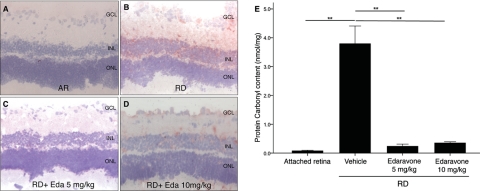
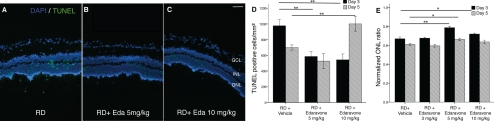
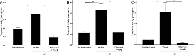
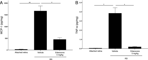
Methods: RD was induced in adult Brown Norway rats by subretinal injection of sodium hyaluronate. Edaravone (3, 5, or 10 mg/kg) or physiologic saline was administered intraperitoneally once a day until death on day 3 or 5. Oxidative stress in the retina was assessed by 4-hydroxynonenal staining or ELISA for protein carbonyl content. Photoreceptor death was assessed by TUNEL and measurement of the outer nuclear layer thickness. Western blot analysis and caspase activity assays were performed. Inflammatory cytokine secretion and inflammatory cell infiltration were evaluated by ELISA and immunostaining, respectively.
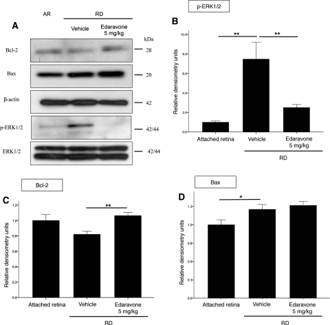
Results: RD resulted in increased generation of ROS. Treatment with 5 mg/kg edaravone significantly reduced the ROS level, along with a decrease in TUNEL-positive cells in the photoreceptor layer. A caspase assay also confirmed decreased activation of caspase-3, -8, and -9 in RD treated with edaravone. The level of the antiapoptotic Bcl-2 was increased in detached retinas after edaravone treatment, whereas the levels of the stress-activated p-ERK1/2 were decreased. In addition, edaravone treatment resulted in a significant decrease in the levels of TNF-α, MCP-1, and macrophage infiltration.
Conclusions: Oxidative stress plays an important role in photoreceptor cell death after RD. Edaravone treatment may aid in preventing photoreceptor cell death after RD by suppressing ROS-induced photoreceptor damage.
Figures

Similar articles
-
Tauroursodeoxycholic acid (TUDCA) protects photoreceptors from cell death after experimental retinal detachment.PLoS One. 2011;6(9):e24245. doi: 10.1371/journal.pone.0024245. Epub 2011 Sep 22. PLoS One. 2011. PMID: 21961034 Free PMC article.
-
Role of oxidative stress in retinal photoreceptor cell death in N-methyl-N-nitrosourea-treated mice.J Pharmacol Sci. 2012;118(3):351-62. doi: 10.1254/jphs.11110fp. Epub 2012 Feb 23. J Pharmacol Sci. 2012. PMID: 22362184
-
Systemic administration of a free radical scavenger, edaravone, protects against light-induced photoreceptor degeneration in the mouse retina.Eur J Pharmacol. 2010 Sep 10;642(1-3):77-85. doi: 10.1016/j.ejphar.2010.05.057. Epub 2010 Jun 8. Eur J Pharmacol. 2010. PMID: 20553915
-
Retinal Diseases Associated with Oxidative Stress and the Effects of a Free Radical Scavenger (Edaravone).Oxid Med Cell Longev. 2017;2017:9208489. doi: 10.1155/2017/9208489. Epub 2017 Jan 18. Oxid Med Cell Longev. 2017. PMID: 28194256 Free PMC article. Review.
-
Edaravone (3-methyl-1-phenyl-2-pyrazolin-5-one), a novel free radical scavenger, for treatment of cardiovascular diseases.Recent Pat Cardiovasc Drug Discov. 2006 Jan;1(1):85-93. doi: 10.2174/157489006775244191. Recent Pat Cardiovasc Drug Discov. 2006. PMID: 18221078 Review.
Cited by
-
Retinal Microvasculature Changes After Repair of Macula-off Retinal Detachment Assessed with Optical Coherence Tomography Angiography.Clin Ophthalmol. 2020 Jun 26;14:1759-1767. doi: 10.2147/OPTH.S214623. eCollection 2020. Clin Ophthalmol. 2020. PMID: 32616995 Free PMC article.
-
Inflammation and Oxidative Stress Gene Variability in Retinal Detachment Patients with and without Proliferative Vitreoretinopathy.Genes (Basel). 2023 Mar 27;14(4):804. doi: 10.3390/genes14040804. Genes (Basel). 2023. PMID: 37107562 Free PMC article.
-
RIPK necrotic cell death pathway in both donor photoreceptor and host immune cells synergize to affect photoreceptor graft survival.FASEB J. 2023 Apr;37(4):e22847. doi: 10.1096/fj.202201137R. FASEB J. 2023. PMID: 36862516 Free PMC article.
-
Inflammatory Mechanisms in the Management and Treatment of Retinal Detachment.Metabolites. 2025 Jul 1;15(7):442. doi: 10.3390/metabo15070442. Metabolites. 2025. PMID: 40710541 Free PMC article. Review.
-
Vitreous Humor Proteome: Targeting Oxidative Stress, Inflammation, and Neurodegeneration in Vitreoretinal Diseases.Antioxidants (Basel). 2022 Mar 6;11(3):505. doi: 10.3390/antiox11030505. Antioxidants (Basel). 2022. PMID: 35326156 Free PMC article. Review.
References
-
- Linsenmeier RA, Padnick-Silver L. Metabolic dependence of photoreceptors on the choroid in the normal and detached retina. Invest Ophthalmol Vis Sci. 2000;41:3117–3123 - PubMed
-
- Zacks DN, Han Y, Zeng Y, Swaroop A. Activation of signaling pathways and stress-response genes in an experimental model of retinal detachment. Invest Ophthalmol Vis Sci. 2006;47:1691–1695 - PubMed
-
- Carmody RJ, McGowan AJ, Cotter TG. Reactive oxygen species as mediators of photoreceptor apoptosis in vitro. Exp Cell Res. 1999;248:520–530 - PubMed
-
- Rotstein NP, Politi LE, German OL, Girotti R. Protective effect of docosahexaenoic acid on oxidative stress-induced apoptosis of retina photoreceptors. Invest Ophthalmol Vis Sci. 2003;44:2252–2259 - PubMed
Publication types
MeSH terms
Substances
Grants and funding
LinkOut - more resources
Full Text Sources
Other Literature Sources
Medical
Research Materials
Miscellaneous
