. 2011 Jun 1;52(6):3784-91.
doi: 10.1167/iovs.10-6386.

A potential role for angiopoietin 2 in the regulation of the blood-retinal barrier in diabetic retinopathy

Affiliations

A potential role for angiopoietin 2 in the regulation of the blood-retinal barrier in diabetic retinopathy

Sampathkumar Rangasamy et al. Invest Ophthalmol Vis Sci. .

Abstract

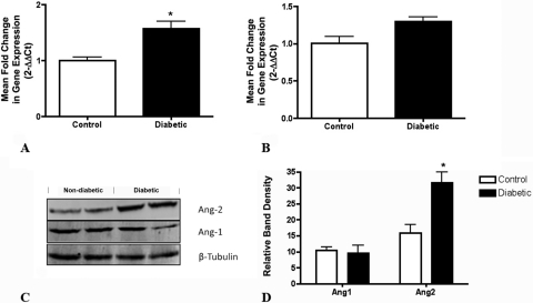
Purpose: Although VEGF has been identified as an important mediator of the blood-retinal barrier alteration in diabetic retinopathy, the hypothesis for this study was that that other molecules, including the angiopoietins (Ang-1 and -2), may play a role. The expression of angiopoietins was analyzed in an animal model of diabetic retinopathy, and the role of Ang-2 in the regulation of diabetes-induced alterations of vascular permeability was characterized.

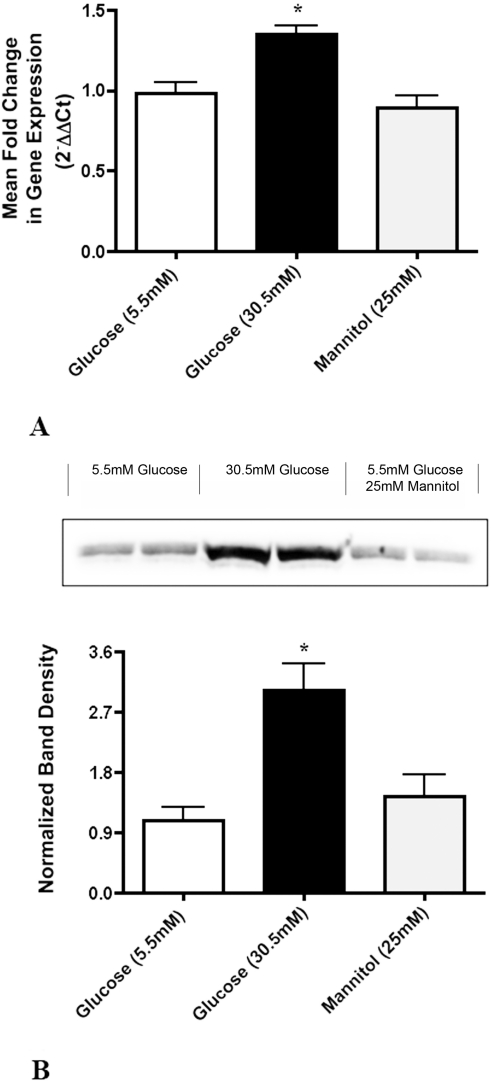
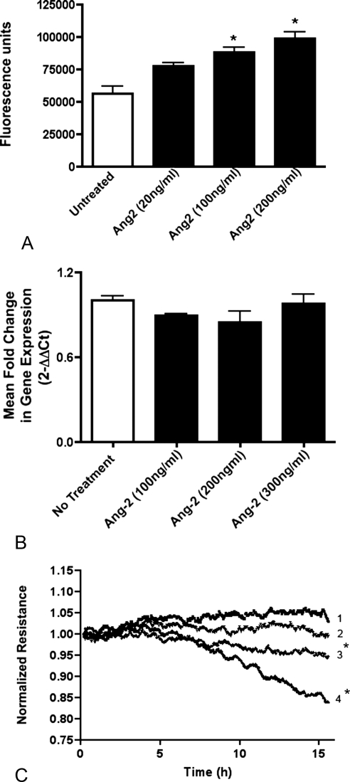
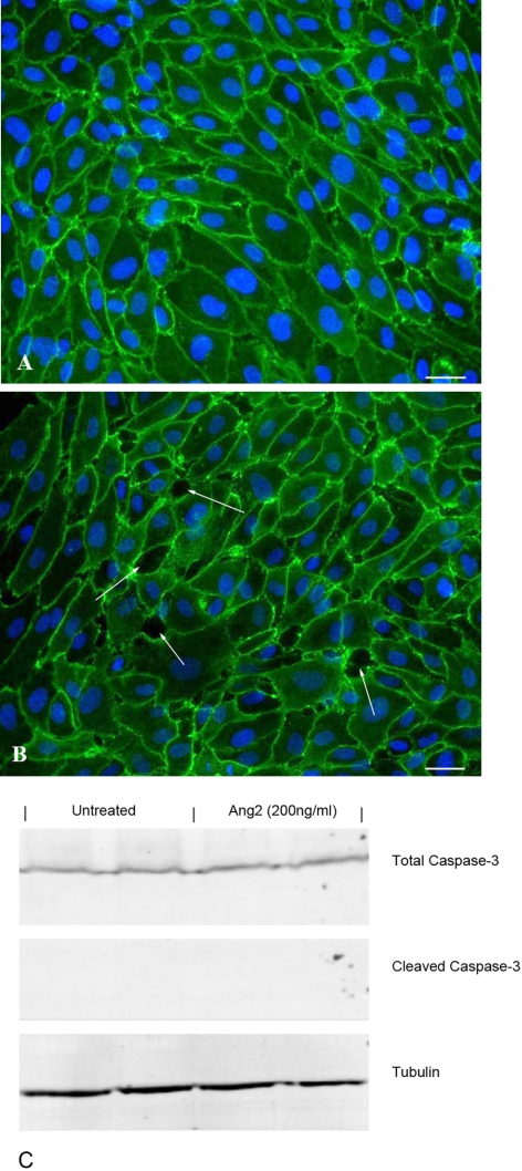
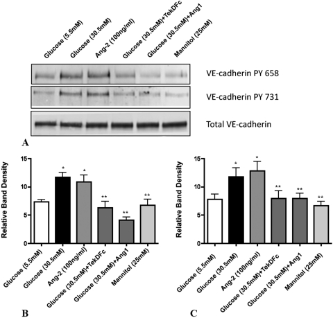
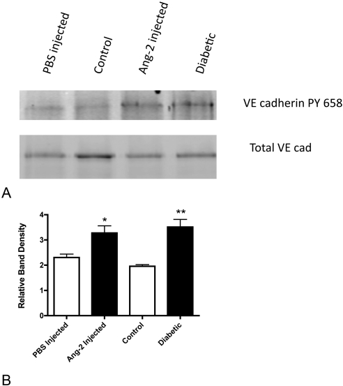
Methods: Diabetes was induced in rats, and human retinal endothelial cells (HRECs) were grown in media with 5.5 or 30.5 mM glucose. Levels of Ang-1 and -2 mRNA and protein were analyzed. Fluorescence-based assays were used to assess the effect of Ang-2 on vascular permeability in vivo and in vitro. The effect of Ang-2 on VE-cadherin function was assessed by measuring the extent of tyrosine phosphorylation.

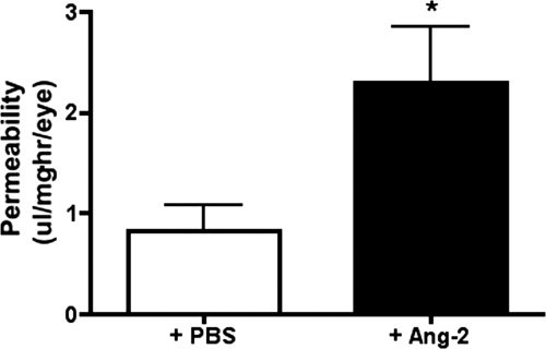
Results: Ang-2 mRNA and protein increased in the retinal tissues after 8 weeks of diabetes and in high-glucose-treated cells. Intravitreal injection of Ang-2 in rats produced a significant increase in retinal vascular permeability. Ang-2 increased HREC monolayer permeability that was associated with a decrease in VE-cadherin and a change in monolayer morphology. High glucose and Ang-2 produced a significant increase in VE-cadherin phosphorylation. CONCLUSIONS; Ang-2 is upregulated in the retina in an animal model of diabetes, and hyperglycemia induces the expression of Ang-2 in isolated retinal endothelial cells. Increased Ang-2 alters VE-cadherin function, leading to increased vascular permeability. Thus, Ang-2 may play an important role in increased vasopermeability in diabetic retinopathy.


Figures

Figure 1.
Figure 1.
Ang-2 was significantly increased in the rat retina after 8 weeks of diabetes. (A) Retinas from diabetic animals (n = 4) demonstrated significantly more Ang-2 mRNA than retinas from nondiabetic controls (n = 6). (B) No significant change in Ang-1 mRNA was detected. *Significantly greater than in the nondiabetic controls (P = 0.031). (C) A representative Western blot demonstrates a significant elevation in Ang-2 expression in the diabetic compared with control retina. (D) Quantitation of Ang-1 and -2 protein levels in control nondiabetic retinas (n = 5) and diabetic retinas (n = 3). *Significantly greater than nondiabetic controls (P = 0.0184). Data are expressed as the mean ± SEM.
Figure 2.
Figure 2.
High glucose increased the expression and secretion of Ang-2 in isolated human retinal microvascular endothelial cells. (A) Ang-2 mRNA levels from cells grown in 30.5 mM glucose was significantly greater than that from cells grown in 5.5 mM glucose or 5.5 mM glucose plus 25 mM mannitol (5.5 mM glucose vs. 30.5 mM glucose, P < 0.01; 25 mM mannitol vs. 30.5 mM glucose, P < 0.05). (B) The relative level of Ang-2 protein in the conditioned medium of cells grown in 5.5 glucose, 30.5 glucose, or 5.5 mM glucose and 25 mM mannitol. Quantitation of band density and normalization to total cell number revealed a significant elevation of Ang-2 protein in the high glucose-treated cells (5.5 mM glucose vs. 30.5 mM glucose, P < 0.01; 25 mM mannitol vs. 30.5 mM glucose, P < 0.05). Data are expressed as the mean ± SEM.
Figure 3.
Figure 3.
Ang-2 alters the endothelial cell barrier. (A) Monolayer permeability to FITC-dextran after Ang-2 treatment. *Significantly greater than untreated cells (untreated versus Ang-2 [20 ng/mL], P > 0.05; untreated versus Ang-2 [100 ng/mL], P < 0.01; untreated versus Ang-2 [200 ng/mL]; P < 0.001). Values represent the mean ± SEM from triplicate wells. (B) Expression of VEGF by human retinal endothelial cells treated with increasing concentrations of Ang-2 for 24 hours. No significant difference was noted at any concentration. (C) Representative ECIS tracing plotted as normalized resistance reflecting the decrease in resistance of endothelial cell monolayers to 100 ng/mL. Ang-2 (tracing 2), 200 ng/mL Ang-2 (tracing 3), 50 ng/mL VEGF (tracing 4) compared with untreated cells (tracing 1). *Significantly less than untreated cells (VEGF versus untreated, P < 0.001; Ang-2 vs. untreated, P < 0.01).
Figure 4.
Figure 4.
Ang-2 induces altered cell surface expression of VE-cadherin and the formation of intercellular gaps in an endothelial cell monolayer. Representative images of VE-cadherin staining in human retinal endothelial cells in untreated (A) or Ang-2-treated (200 ng/mL) (B) conditions. A disruption of the continuous pattern of VE-cadherin staining along cell borders and the presence of open gaps between adjacent cells (arrows) is seen in the Ang-2 treated cultures. (C) Representative Western blot of cleaved and full-length caspase-3 levels in control cells and cells treated with Ang-2. β-tubulin was used as a loading control. Bar, 10 μm.
Figure 5.
Figure 5.
Intraocular injection of purified Ang-2 increased retinal vascular permeability. Normal nondiabetic rats receiving an intravitreal injection of purified Ang-2 (100 ng) show a nearly threefold increase in the extravascular accumulation of FITC-BSA in the retinal tissues compared to animals receiving an injection of PBS alone (2.291 ± 0.556, n = 4 vs. 0.831 ± 0.254, n = 4). *Significantly different, P = 0.014.
Figure 6.
Figure 6.
High glucose and Ang-2 induce VE-cadherin phosphorylation in vitro. (A) Representative Western blot image of phospho-VE-cadherin in human retinal endothelial cells. Quantitation of band density for P-Y658 (B) and P-Y731 (C) relative to total VE-cadherin for the indicated treatment conditions. *Significantly greater than 5.5 mM glucose. **Significantly less than 30.5 mM glucose alone.
Figure 7.
Figure 7.
Increased VE-cadherin phosphorylation in the retina in response to Ang-2 and diabetes. (A) Representative Western blot image of phospho-VE-cadherin in rat retina extracts. Quantitation of band density for P-Y658 relative to total VE-cadherin for the indicated experimental conditions. *Significantly greater than in the PBS injected animals (P = 0.04). **Significantly greater than in the control nondiabetic animals (P = 0.01).

References

    1. Congdon NG, Friedman DS, Lietman T. Important causes of visual impairment in the world today. JAMA. 2003;290:2057–2060 - PubMed
    1. Geiger B, Ayalon O. Cadherins Ann Rev Cell Biol. 1992;8:307–333 - PubMed
    1. Aiello LP. Angiogenic pathways in diabetic retinopathy. N Engl J Med. 2005;353:839–841 - PubMed
    1. Adamis AP. Is diabetic retinopathy an inflammatory disease? Br J Ophthalmol. 2002;86:363–365 - PMC - PubMed
    1. Biester S, Ziemssen F, Ulrich Bartz-Schmidt K, Gelisken F. Is intravitreal bevacizumab treatment effective in diffuse diabetic macular edema? Graefes Arch Clin Exp Ophthalmol. 2009;247:1575–1577 - PubMed

Publication types

MeSH terms