. 2011 Mar 4;331(6021):1203-7.
doi: 10.1126/science.1201730. Epub 2011 Feb 10.

Different B cell populations mediate early and late memory during an endogenous immune response

Affiliations

Different B cell populations mediate early and late memory during an endogenous immune response

Kathryn A Pape et al. Science. .

Abstract

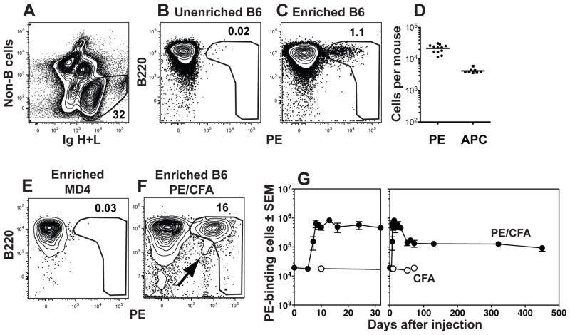
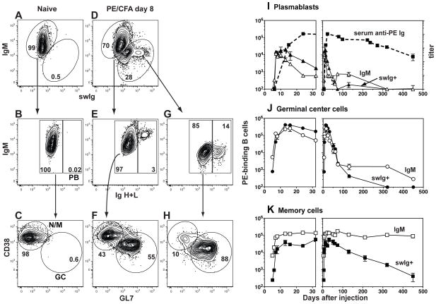
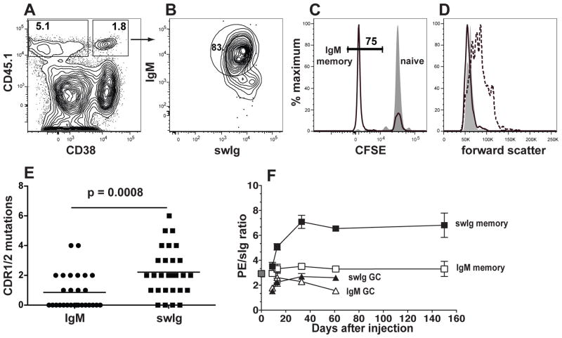
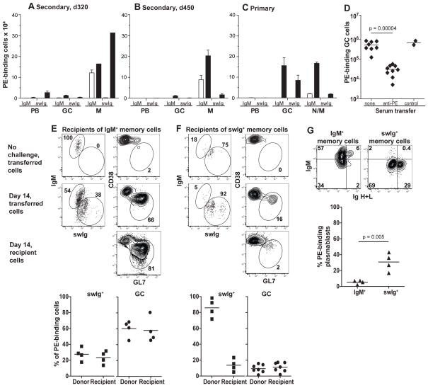
Memory B cells formed in response to microbial antigens provide immunity to later infections; however, the inability to detect rare endogenous antigen-specific cells limits current understanding of this process. Using an antigen-based technique to enrich these cells, we found that immunization with a model protein generated B memory cells that expressed isotype-switched immunoglobulins (swIg) or retained IgM. The more numerous IgM(+) cells were longer lived than the swIg(+) cells. However, swIg(+) memory cells dominated the secondary response because of the capacity to become activated in the presence of neutralizing serum immunoglobulin. Thus, we propose that memory relies on swIg(+) cells until they disappear and serum immunoglobulin falls to a low level, in which case memory resides with durable IgM(+) reserves.


Figures

Fig. 1
Fig. 1
Detection of PE-binding B cells. (A) B cells were identified by flow cytometry in spleen and lymph node samples as cells that did not bind a cocktail of antibodies specific for CD4, CD8, CD11c, Gr1, or F480 (non-B cells) and expressed Ig heavy and light chains (H+L, both intracellular and extracellular). The cells with large amounts of Ig are plasmablasts. (B) Representative flow cytometric analysis of unenriched spleen and lymph node B cells from a naïve B6 mouse after staining with PE. (C) Representative flow cytometric analysis of spleen and lymph node B cells from a naïve B6 mouse after staining with PE and enrichment with anti-PE magnetic beads. (D) Number of PE- or allophycocyanin (APC)-specific B cells in individual naïve B6 mice. The bar indicates the mean. (E–F) Representative flow cytometric analysis of spleen and lymph node B cells from a naïve MD4 Rag1−/− mouse (E) or a B6 mouse, 24 days after subcutaneous injection of 15 μg PE and CFA (F), that were stained with PE and enriched with anti-PE magnetic beads. The PE gate was drawn to exclude cells that bind antigen-antibody complexes (arrow in F). (G) Combined data from multiple experiments showing the numbers of PE-binding B cells in the spleen and lymph nodes at the indicated times after subcutaneous injection of 15 μg PE and CFA (filled circles) or CFA alone (open circles) shown over the first 32 (left panel) or 450 (right panel) days. The mean and SEM (N=3–6) is shown for all time points except the day 61 time point for PE and CFA injected mice, where (N=2) and the mean is shown without error bars, and the day 52 and day 72 time points for CFA alone injected mice (N=1). (27)
Fig. 2
Fig. 2
Detection of PE-specific plasmablasts, germinal center cells, and memory B cells. PE-binding cells enriched from the spleen and lymph nodes of (A–C) a naive B6 mouse or (D–H) a B6 mouse on day 8 after subcutaneous injection of PE and CFA, identified as shown in Fig. 1. (A and D) Flow cytometry based gating strategy used to identify cells expressing surface IgM and/or surface and intracellular IgG1, IgG2c, IgG2b, IgG3, IgA, and IgE. (B, E and G) Flow cytometry based gating strategy used to identify Iglow non-plasmablasts (left) or Ighigh plasmablasts (PB) (right) within the IgM+ or swIg+ populations. (C, F and H) Flow cytometry based gating strategy used to identify CD38+ GL7 naive/memory or CD38 GL7+ germinal center (GC) cells within the non-PB populations. (I–K) Combined data from multiple experiments showing the numbers of IgM+ (open symbols, solid line) or swIg+ (filled symbols, solid line) PE-binding plasmablasts (I), germinal center cells (J), or memory cells (K) in the spleen and lymph nodes after subcutaneous injection of PE and CFA on day 0. The mean and SEM (N=3–6) is shown for all time points except days 5, 7, 24, 61, and 74 where (N=2) and the mean is shown without error bars. The mean titer and SEM (N=4–10) of total PE-specific Ig in the serum is shown (dashed line) in (I). (27)
Fig. 3
Fig. 3
Properties of the PE-specific IgM+ memory B cell population. (A–D) PE-binding cells enriched from B6 recipients of CD45.1+ CFSE-labeled B cells, 9 days after subcutaneous injection of PE and CFA or CFA alone identified as shown in Fig. 1. (A) Flow cytometry based gating strategy used to identify CD45.1+ donor PE-specific B cells that did (right) or did not (left) express CD38. (B) Flow cytometry based gating strategy used to identify CD38+,CD45.1+ PE-specific B cells that expressed IgM. (C) Flow cytometric analysis of CFSE expression in CD45.1+ PE-specific CD38+ IgM+ cells from mice injected with CFA alone (filled) or PE and CFA (black line). The frequency of CFSElow cells is indicated for PE and CFA injected mice. (D) Representative flow cytometric analysis of forward light scatter of PE-specific IgM+ CD38+ naïve cells from mice injected with CFA alone (filled), or PE-specific CD38+ IgM+ memory cells (black line), or CD38 IgM+ cells from mice injected with PE and CFA (dashed line). (E) The number of mutations resulting in amino acid substitutions, in the CDR1 and 2 VH genes for each of 28 PE-binding IgM+ or IgG1+ clones from mice injected 25 days earlier with PE and CFA. Statistical significance was established with an unpaired Student’s t-test. (F) Combined data from two independent experiments showing the ratio of PE fluorescence intensity divided by total Ig fluorescence intensity for naive (grey filled square) IgM+ memory (open squares), swIg+ memory (black filled squares), IgM+ germinal center (open triangles), or swIg+ germinal center (black filled triangles) cells at the indicated times after PE and CFA injection. The mean and SEM (N=3–4) is shown for all time points except day 0 and day 61 where (N=2) and the mean is shown without error bars. (27)
Fig. 4
Fig. 4
Functional capabilities of memory B cells. (A–D) PE-binding cells enriched from the spleen and lymph nodes of B6 mice and identified as shown in Figs. 1 and 2. (A–C) Closed bars represent the numbers of plasmablasts on day 4 and germinal center cells or memory cells on day 14 after intraperitoneal injection of PE and CFA into day 320 memory (A), day 450 memory (B), or naïve (C) mice. Open bars represent numbers before the intraperitoneal challenge injection. The mean and SEM (N=3–4) is shown for all groups except for the day 320 memory cells after challenge where (N=2) and the mean is shown without error bars (range=102,678). (D) Numbers of PE-binding germinal centers present on day 12–14 in mice left untreated, or injected with serum from mice immunized with PE or control antigens (chicken ovalbumin or allophycocyanin) on days 3–6 after antigen injection. Statistical significance was established with an unpaired Student’s t-test. (E–F) Flow cytometric analysis of PE-specific cells of donor or recipient origin in the spleen and lymph nodes of CD45.1+ recipients of IgM+ (E) or swIg+ (F) CD45.2+ memory cells before and 10–14 days after PE and CFA challenge. Gates used to identify IgM+, swIg+, and germinal center cells are shown. Scatterplots show the percentage of the indicated cell types present in the PE-binding populations of donor or recipient origin in individual mice 10–14 days after PE and CFA challenge. (G) PE-specific cells derived from transferred IgM+ or swIg+ memory cells in individual recipient mice 5 days after PE and CFA challenge. Plasmablasts were identified as Ighigh cells. Statistical significance was established with an unpaired Student’s t-test. (27)

Comment in

References

    1. McHeyzer-Williams LJ, McHeyzer-Williams MG. Annu Rev Immunol. 2005;23:487. - PubMed
    1. Tarlinton DM. Immunol Cell Biol. 2008;86:133. - PubMed
    1. Maul RW, Gearhart PJ. Adv Immunol. 2010;105:159. - PMC - PubMed
    1. Yoshida T, et al. Immunol Rev. 2010;237:117. - PubMed
    1. Tangye SG, Good KL. J Immunol. 2007;179:13. - PubMed

Publication types

MeSH terms