. 2011 Feb 11;331(6018):775-8.
doi: 10.1126/science.1199326.

Leishmania RNA virus controls the severity of mucocutaneous leishmaniasis

Affiliations

Leishmania RNA virus controls the severity of mucocutaneous leishmaniasis

Annette Ives et al. Science. .

Abstract

Mucocutaneous leishmaniasis is caused by infections with intracellular parasites of the Leishmania Viannia subgenus, including Leishmania guyanensis. The pathology develops after parasite dissemination to nasopharyngeal tissues, where destructive metastatic lesions form with chronic inflammation. Currently, the mechanisms involved in lesion development are poorly understood. Here we show that metastasizing parasites have a high Leishmania RNA virus-1 (LRV1) burden that is recognized by the host Toll-like receptor 3 (TLR3) to induce proinflammatory cytokines and chemokines. Paradoxically, these TLR3-mediated immune responses rendered mice more susceptible to infection, and the animals developed an increased footpad swelling and parasitemia. Thus, LRV1 in the metastasizing parasites subverted the host immune response to Leishmania and promoted parasite persistence.


Figures
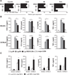
Fig. 1
Fig. 1
Metastasizing L.g. parasites activate bone-marrow macrophages to elevate proinflammatory cytokine and chemokine levels. (A) Transcript and (B and C) secreted protein levels induced after C57BL/6 or BALB/c macrophage infection (ratio 1:10) with Leishmania parasites [two L.g.M− clones (Lg03 and Lg17); two L.g.M+ clones (Lg13 and Lg21); L.g.M5313(M+); L.g. derived from h-MCL (−L.g.1398) or hCL (L.g.1881) lesions; and L.major LV39] for 6 hours. Results were confirmed in several independent experiments (n > 3), and data reflect mean ± SD transcript or protein increase relative to unstimulated controls. Significance was determined at *P ≤ 0.05, and **P ≤ 0.01 for L.g.M+ or h-MCL versus L.g.M−, h-CL, and/or L. major LV39-stimulated macrophages.
Fig. 2
Fig. 2
L.g.M+ or h-MCL parasite-dependent induction of IFN-β and proinflammatory mediators by macrophages uses TLR3 and TRIF. (A and C) Secreted protein and (B) transcript levels of cytokines and chemokines induced after infection of macrophages (ratio 1:10) with Leishmania parasites [two L.g.M+ clones (Lg13 and Lg21), two L.g.M− clones (Lg03 and Lg17), and L.g.M5313(M+)] for 6 and 2 hours, respectively. Results were confirmed in several independent experiments (n = 3), and data reflect mean ± SD transcript or protein increase relative to unstimulated controls of L.g.M+ or L.g.M−. Significance was determined between C57BL/6 and deficient macrophages (A and C) or between L.g.M+ or h-MCL and L.g M− and h-CL parasites (B) at *P ≤ 0.05 and **P ≤ 0.01. n.i, not induced.
Fig. 3
Fig. 3
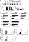
High LRV1 burden within metastasizing L.g. promastigotes stimulates cytokine and chemokine production in macrophages via TLR3. (A) ssRNAse-treated nucleic acids were DNAse treated, and the 5.3-kb LRV1 dsRNA band visualized by gel electrophoresis. (B) LRV1 virus burden within Leishmania parasites was assessed by qRT-PCR with LRV1 and Leishmania Kmp11 gene primers; significance was determined between metastasizing (L.g.M+ and h-MCL) versus nonmetastasizing (L.g.M− and h-CL) parasites. (C) Nucleic acids from L.g.M5313(M+) promastigotes, pretreated with a ssRNA-specific RNAse, were treated with DNAse or with the dsRNA-specific RNAse III and separated by gel electrophoresis with the marker Lambda-DNA–Eco RI + Hind III (HindIII). (D) Macrophages were stimulated with purified LRV1 dsRNA (1 µg/ml) in endotoxin-free (LAL) water, poly(I:C) (1 µg/ml), or lipopolysaccharide (LPS, 100 ng/ml) for 4 hours. Transcript levels were assessed relative to unstimulated C57BL/6 macrophages by qRT-PCR. Results are expressed as mean ± SD (n=2). (E) Protein abundance was quantified after infection of macrophages (ratio 1:10) with L.g.M4147–LRVhigh (clones 2 and 3) or L.g.M4147–LRVneg (clones 3 and 4) parasites after 6 hours. Controls: L.g.M− (Lg17), L.g.M5313(M+), poly(I:C) (2 µg/ml), and LPS (100 ng/ml). Data reflect mean ± SD of protein secretion relative to unstimulated controls (n = 2). Significance was determined at *P ≤ 0.05 or **P ≤ 0.01.
Fig. 4
Fig. 4
TLR3−/− mice infected with L.g.M+ parasites have decreased disease pathology when compared to wild-type C57BL/6. Footpads (n ≥ 4) were infected with 3 × 10⁶ parasites. (A) Footpad swelling was measured weekly and (B) parasite burden (n=3) was determined at 4 weeks after infection by qRT-PCR with Leishmania Kmp11 gene-specific primers. Representative data of two experiments, expressed as mean ± SEM of all mice infected per group, with statistical significance at *P ≤ 0.05 and **P ≤ 0.01.

References

    1. Weigle K, Saravia NG. Clin. Dermatol. 1996;14:433. - PubMed
    1. Vergel C, et al. J. Infect. Dis. 2006;194:503. - PubMed
    1. Martinez JE, Alba L. Trans. R. Soc. Trop. Med. Hyg. 1992;86:392. - PubMed
    1. Barral A, et al. Am. J. Pathol. 1995;147:947. - PMC - PubMed
    1. Amato VS, Tuon FF, Bacha HA, Neto VA, Nicodemo AC. Acta Trop. 2008;105:1. - PubMed

Publication types

MeSH terms

Associated data