. 2011 Apr 1;108(7):837-46.
doi: 10.1161/CIRCRESAHA.110.232306. Epub 2011 Feb 10.

Mitochondrial oxidative stress mediates angiotensin II-induced cardiac hypertrophy and Galphaq overexpression-induced heart failure

Affiliations

Mitochondrial oxidative stress mediates angiotensin II-induced cardiac hypertrophy and Galphaq overexpression-induced heart failure

Dao-Fu Dai et al. Circ Res. .

Abstract

Rationale: Mitochondrial dysfunction has been implicated in several cardiovascular diseases; however, the roles of mitochondrial oxidative stress and DNA damage in hypertensive cardiomyopathy are not well understood.

Objective: We evaluated the contribution of mitochondrial reactive oxygen species (ROS) to cardiac hypertrophy and failure by using genetic mouse models overexpressing catalase targeted to mitochondria and to peroxisomes.

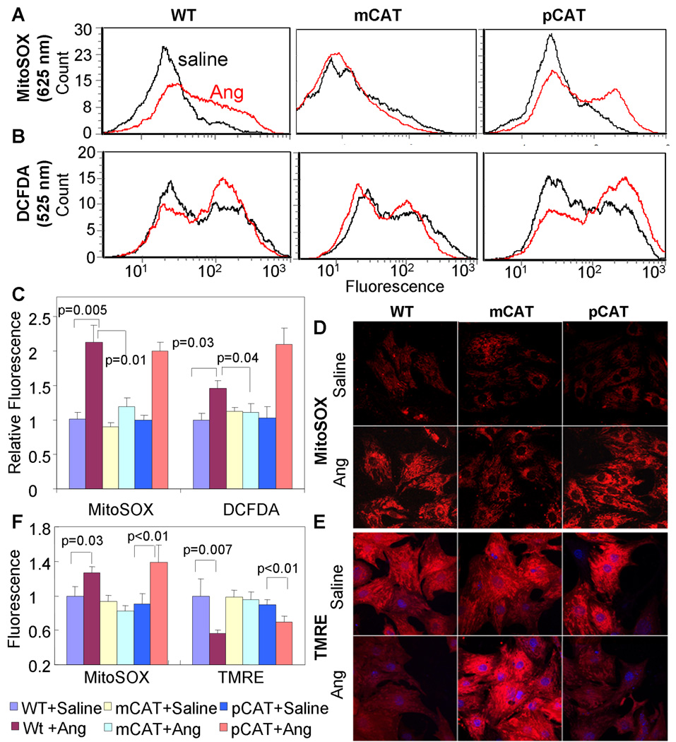
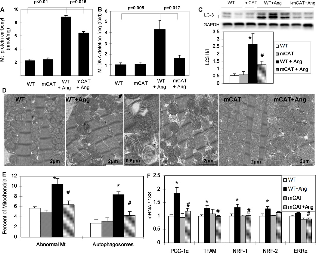
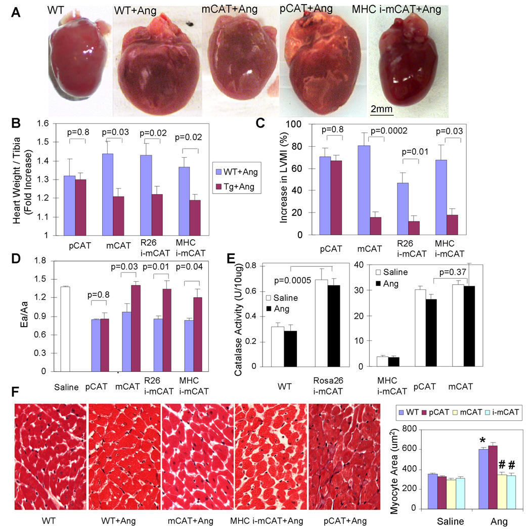
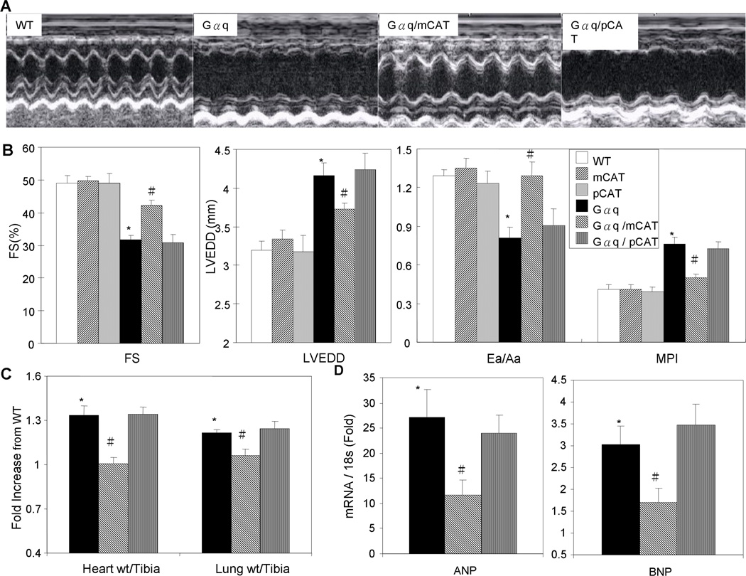
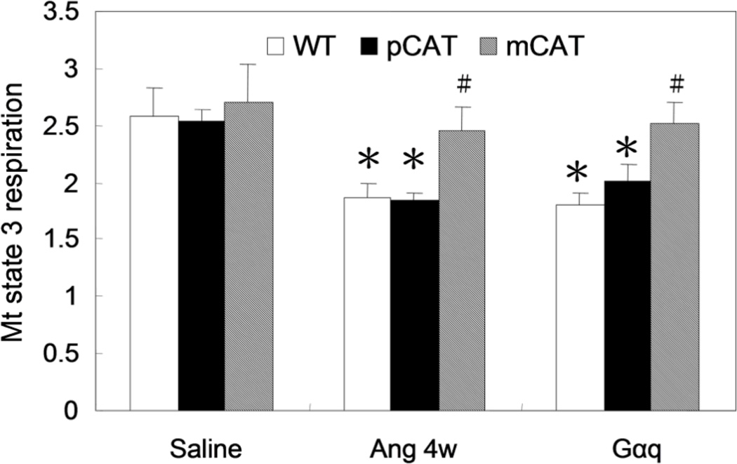
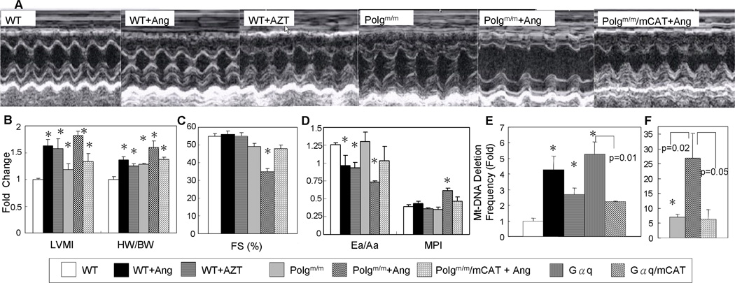
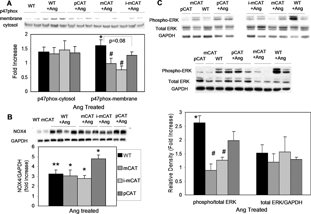
Methods and results: Angiotensin II increases mitochondrial ROS in cardiomyocytes, concomitant with increased mitochondrial protein carbonyls, mitochondrial DNA deletions, increased autophagy and signaling for mitochondrial biogenesis in hearts of angiotensin II-treated mice. The causal role of mitochondrial ROS in angiotensin II-induced cardiomyopathy is shown by the observation that mice that overexpress catalase targeted to mitochondria, but not mice that overexpress wild-type peroxisomal catalase, are resistant to cardiac hypertrophy, fibrosis and mitochondrial damage induced by angiotensin II, as well as heart failure induced by overexpression of Gαq. Furthermore, primary damage to mitochondrial DNA, induced by zidovudine administration or homozygous mutation of mitochondrial polymerase γ, is also shown to contribute directly to the development of cardiac hypertrophy, fibrosis and failure.

Conclusions: These data indicate the critical role of mitochondrial ROS in cardiac hypertrophy and failure and support the potential use of mitochondrial-targeted antioxidants for prevention and treatment of hypertensive cardiomyopathy.


Conflict of interest statement

Disclosures: No conflicts to disclose

Figures

Fig. 1
Fig. 1
Angiotensin-induced mitochondrial oxidative stress and reduction of mitochondrial membrane potential in neonatal mouse cardiomyocytes. Ang (1µM) induced a significant increase in mitochondrial ROS (MitoSOX, A) and total cellular ROS (DCFDA,B) measured by flow cytometry in WT cardiomyocytes (left column A–B). Ang-induced mitochondrial (A) and total ROS (B) is substantially inhibited in mCAT (middle column) but not pCAT (right column) cardiomyocytes, summarized in panel C (mean ± SEM of histograms of triplicate samples). Confocal microscopy shows similar results obtained with MitoSOX (D) and demonstrates that mitochondrial membrane potential, indicated by TMRE fluorescence (E), is reduced in WT cardiomyocytes treated with Ang. Both mitochondrial ROS (MitoSOX) and mitochondrial membrane potential (TMRE) are protected in mCAT but not pCAT cardiomyocytes (middle and right panels of D and E, respectively). Confocal images are quantitated in panel F (see supplemental methods).
Fig. 2
Fig. 2
Ang (1.1mg/kg/d) for 4 weeks induced cardiac mitochondrial damage, autophagy and biogenesis through mitochondrial ROS. (A) Protein carbonyl content was quantified using a DNPH-based enzyme immunoassay of cardiac mitochondrial protein extracts. Ang significantly increased cardiac mt-protein carbonylation, which was significantly protected in mCAT mice, n=9–10 (B) Mitochondrial DNA deletion frequencies quantified by the random mutation assay showed a greater than 4-fold increase after Ang, and this was substantially attenuated in mCAT mouse hearts, n=8–10 (C) Western blots showed that Ang significantly increased the LC-3 II/I ratio, which was significantly attenuated by MHC-i-mCAT. (D) Transmission electron microscopy showed damaged mitochondria (arrowheads) and autophagosomes/autolysosome (arrows) (magnification as indicated). (E) Quantitative analysis of electron micrographs showed that Ang significantly increased the proportion of damaged WT mitochondria (see Methods for definition), as well as the number of autophagosomes/autolysosomes that appear to contain damaged mitochondria. Both of these findings were significantly attenuated in Ang-treated mCAT mouse hearts. (F) Expression of genes implicated in mitochondrial biogenesis was significantly increased in WT hearts after Ang, but was substantially attenuated in Ang-treated mCAT mouse hearts. *p<0.05 for WT vs. WT+Ang, #p<0.05 for WT+Ang vs. mCAT+Ang, n=6–12.
Fig. 3
Fig. 3
Ang-induced cardiac hypertrophy was protected in mice overexpressing mCAT but not pCAT. As seen by heart dimension (A), heart weight normalized to tibia length (B) and LVMI(C), Ang resulted in significant cardiac hypertrophy in WT littermates of each genotype (blue bars). (D) Ea/Aa is normally > 1, as seen in WT treated with saline, but Ang results in diastolic dysfunction in WT (blue bars), shown by Ea/Aa <1. In Panels A–D all Ang-induced changes are significantly attenuated in mice with either constitutive or inducible overexpression of mCAT, but not by overexpression of pCAT (red bars). (E) Total cellular catalase activity (per mg cardiac protein by Amplex Red assay) showed activities for R26-i-mCAT, MHC-i-mCAT, pCAT and mCAT that were 2.2, 12.2, 95.4 and 101.8 fold greater than WT, respectively. There was no significant difference in catalase activity after Ang treatment (black bars). (F) Cardiomyocytes cross-sectional area significantly increased after Ang, which was attenuated in mCAT but not pCAT. Normalized to saline-treated WT littermates of each group (See Online Tables II–III for full data). *p<0.01 compared with saline treated groups. # p<0.01 compared with Ang- treated WT.
Fig. 4
Fig. 4
Gαq overexpression caused heart failure was protected in mCAT but not pCAT mice. (A) Echocardiography showed that 16-week-old Gαq heterozygous mice had poor contractility and enlargement of the LV chamber. The heart failure phenotypes in Gαq mice were substantially attenuated by overexpression of mCAT but not pCAT. (B) Quantitative analysis of echocardiography demonstrated that compared to WT mice, Gαq mice had significantly lower fractional shortening (FS), larger LV end-diastolic dimension (LVEDD), lower Ea/Aa by tissue Doppler and worsening of the MPI. All of these changes were significantly attenuated in compound heterozygous Gαq/mCAT mice but not Gαq/pCAT mice. (C) Relative to WT littermates, both normalized heart weight and lung weight were significantly increased in Gαq and Gαq/pCAT mice, but were substantially protected in Gαq/mCAT mice. (D) Relative to WT littermates, expression of ANP and BNP genes (normalized to 18S) was significantly upregulated in Gαq and Gαq/pCAT mice, but this increase was significantly less in Gαq/mCAT mice. *p<0.05 for WT vs. Gαq, #p<0.05 for Gαq vs. Gαq/mCAT, n= 5–10/group.
Fig. 5
Fig. 5
Mitochondrial state 3 respiration was measured using saponin-permeabilized cardiac muscle fibers from the LV apex, in the presence of excess pyruvate/ malate, stimulated by ADP, and normalized to tissue weight (nmol O2/min/mg). The state 3 respiratory capacity in WT mice significantly declined by 28% after 4 weeks of Ang II, and this was protected in mCAT but not pCAT mice. Gαq overexpression caused a 30% reduction of maximal respiratory capacity and this was protected in Gαq/mCAT but not Gαq/pCAT mice. *p<0.05 compared with WT, #p<0.05 for mCAT+Ang vs. WT+Ang and Gαq/mCAT vs. Gαq, n=4–6/group.
Fig. 6
Fig. 6
Mitochondrial DNA damage can induce cardiac hypertrophy and failure. (A) Echocardiography demonstrated that both Ang and AZT (zidovudine) treatment of WT mice induced LV hypertrophy with preserved systolic function. In contrast, Ang treatment of Polgm/m mice caused significant impairment of LV contractility and severe hypertrophy, which is ameliorated by mCAT. (B) Relative to untreated WT mice, both echocardiographic LVMI and normalized heart weight increased significantly after Ang or AZT. These parameters of cardiac hypertrophy increased even more dramatically when Polgm/m mice were treated with Ang. WT mice treated with Ang or AZT had preserved systolic function measured by FS (C) and a slight decline in diastolic function (Ea/Aa) with no change in MPI (D). Polgm/m mice showed normal systolic and diastolic function, but Polgm/m mice treated with Ang developed heart failure with worsening of myocardial performance and impairment of both systolic and diastolic function, all of which were attenuated by mCAT (C–D). (E) Mutation assay showed that Ang, AZT and Gαq resulted in significantly increased mtDNA deletion frequencies: 4.3, 2.7 and 5.2-fold above WT, respectively. Expression of mCAT in Gαq mice, which attenuated heart failure (Figures 4–5), also significantly decreased the mtDNA deletion frequency. Polgm/m mice have a 7-fold increase in mtDNA deletion frequency, which rises to 26.9-fold after Ang treatment and this is alleviated by mCAT. *p<0.05 compared with WT. n=6–10 for echocardiography, n=8–12 for deletion assay.
Fig. 7
Fig. 7
(A) Ang increased p47phox membrane translocation, which was attenuated by mCAT (p<0.05) and pCAT (p=0.08). (B) Ang significantly increased NOX4 expression (p<0.01), which was not altered by mCAT or pCAT. (C) Ang-induced phosphorylation of ERK1/2 is significantly reduced in mCAT but not pCAT mice. *p<0.05 for Ang-treated vs. saline treated; #p<0.05 compared with WT; n=5–7.

References

    1. Lifton RP, Gharavi AG, Geller DS. Molecular mechanisms of human hypertension. Cell. 2001;104:545–556. - PubMed
    1. Gardin JM, Lauer MS. Left ventricular hypertrophy: the next treatable, silent killer? Jama. 2004;292:2396–2398. - PubMed
    1. Mollnau H, Wendt M, Szocs K, Lassegue B, Schulz E, Oelze M, Li H, Bodenschatz M, August M, Kleschyov AL, Tsilimingas N, Walter U, Forstermann U, Meinertz T, Griendling K, Munzel T. Effects of angiotensin II infusion on the expression and function of NAD(P)H oxidase and components of nitric oxide/cGMP signaling. Circ Res. 2002;90:E58–E65. - PubMed
    1. Doughan AK, Harrison DG, Dikalov SI. Molecular mechanisms of angiotensin II-mediated mitochondrial dysfunction: linking mitochondrial oxidative damage and vascular endothelial dysfunction. Circ Res. 2008;102:488–496. - PubMed
    1. Kimura S, Zhang GX, Nishiyama A, Shokoji T, Yao L, Fan YY, Rahman M, Abe Y. Mitochondria-derived reactive oxygen species and vascular MAP kinases: comparison of angiotensin II and diazoxide. Hypertension. 2005;45:438–444. - PubMed

Publication types

MeSH terms