Review
. 2011 Apr;18(4):571-80.
doi: 10.1038/cdd.2010.191. Epub 2011 Feb 11.

The Beclin 1 network regulates autophagy and apoptosis

Affiliations
Review

The Beclin 1 network regulates autophagy and apoptosis

R Kang et al. Cell Death Differ. 2011 Apr.

Abstract

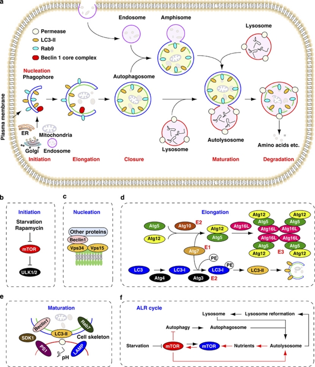
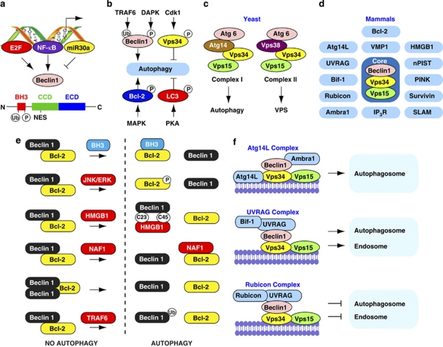
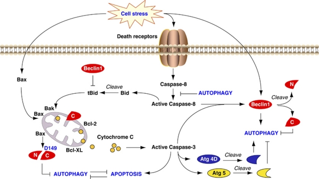
Beclin 1, the mammalian orthologue of yeast Atg6, has a central role in autophagy, a process of programmed cell survival, which is increased during periods of cell stress and extinguished during the cell cycle. It interacts with several cofactors (Atg14L, UVRAG, Bif-1, Rubicon, Ambra1, HMGB1, nPIST, VMP1, SLAM, IP(3)R, PINK and survivin) to regulate the lipid kinase Vps-34 protein and promote formation of Beclin 1-Vps34-Vps15 core complexes, thereby inducing autophagy. In contrast, the BH3 domain of Beclin 1 is bound to, and inhibited by Bcl-2 or Bcl-XL. This interaction can be disrupted by phosphorylation of Bcl-2 and Beclin 1, or ubiquitination of Beclin 1. Interestingly, caspase-mediated cleavage of Beclin 1 promotes crosstalk between apoptosis and autophagy. Beclin 1 dysfunction has been implicated in many disorders, including cancer and neurodegeneration. Here, we summarize new findings regarding the organization and function of the Beclin 1 network in cellular homeostasis, focusing on the cross-regulation between apoptosis and autophagy.


Figures

Figure 1
Figure 1
Stages of autophagy. (a) Different types of autophagy. LC3-II is a marker of Atg5/Atg7-dependent autophagy, whereas Rab-9 is a marker of Atg5/Atg7-independent autophagy. (b) The initiation is sustained by activation of ULK1 and ULK2 complexes, which are inhibited by mTOR. (c) The nucleation depends on Beclin 1-Vps34-Vps15 core complexes and other proteins. (d) The elongation of the phagophore is mediated by two ubiquitin-like conjugation systems that together promote the assembly of the ATG16L complex and the processing of LC3. PE, phosphatidylethanolamine. (e) The maturation is promoted by LC3, Beclin 1, the lysosomal membrane proteins LAMP-1 and LAMP-2, the GTP-binding protein RAB7, the ATPase SKD1, the cell skeleton, the pH of lysosomes and possibly presenilin 1 (PS1). (f) Autophagic lysosome reformation (ALR) cycle. mTOR signaling is inhibited during initiation of autophagy, but reactivated by prolonged starvation. Reactivation of mTOR is autophagy-dependent and requires the degradation of autolysosomal products. Increased mTOR activity attenuates autophagy and generates proto-lysosomal tubules and vesicles that extrude from autolysosomes and ultimately mature into functional lysosomes, thereby restoring the full complement of lysosomes in the cell (figure modified from , , , )
Figure 2
Figure 2
The complex Beclin 1 network. (a) NF-κB and E2F positively regulates Beclin 1 expression, whereas miR30a negatively regulates Beclin 1 expression. Conversely, miR-9 increases autophagy by regulating histone deacetylases activity in lymphoma cells (not shown). Structurally, Beclin 1 has a BH3 domain, a central CCD and an ECD. (b) Phosphorylation and ubiquitination modification regulates autophagy. (c) In yeast, Atg6-Vps34-Vps15 complexes I and II regulate autophagy and Vps, respectively. (d) In mammals, Beclin 1-Vps34-Vps15 core complexes and Beclin 1's binding proteins. (e) Models of Beclin 1–Bcl-2 complex dissociation in autophagy. (f) Model of the role of the three Beclin 1-Vps34-Vps15 complexes. The Atg14L complex functions positively in autophagosome formation. The UVRAG complex functions positively in autophagosome and endosome maturation. The Rubicon complex functions negatively in autophagosome and endosome maturation (figure modified from , )
Figure 3
Figure 3
Crosstalk between apoptosis and autophagy. Autophagy and apoptosis share common stimuli and signaling pathways, and exhibit some degree of mutual inhibition. During sustained exposure to apoptotic stimuli, caspase-mediated cleavage of Beclin 1 generates fragments (‘N' and ‘C') that lose their ability to induce autophagy. The C-terminal fragment translocates to the mitochondria and sensitizes cells to apoptotic signals. Although apoptosis-associated cleavage of Beclin 1 and Atg5 inactivates autophagy, the cleavage of Atg4D by caspase-3 generates a fragment with increased autophagy activity. Moreover, autophagy inhibits apoptosis partly by degrading active caspase-8 or preventing activation of Bid by Beclin 1

References

    1. Yang Z, Klionsky DJ. Eaten alive: a history of macroautophagy. Nat Cell Biol. 2010;12:814–822. - PMC - PubMed
    1. Tooze SA, Yoshimori T. The origin of the autophagosomal membrane. Nat Cell Biol. 2010;12:831–835. - PubMed
    1. Nishida Y, Arakawa S, Fujitani K, Yamaguchi H, Mizuta T, Kanaseki T, et al. Discovery of Atg5/Atg7-independent alternative macroautophagy. Nature. 2009;461:654–658. - PubMed
    1. Zhang J, Randall MS, Loyd MR, Dorsey FC, Kundu M, Cleveland JL, et al. Mitochondrial clearance is regulated by Atg7-dependent and -independent mechanisms during reticulocyte maturation. Blood. 2009;114:157–164. - PMC - PubMed
    1. Scarlatti F, Maffei R, Beau I, Codogno P, Ghidoni R. Role of non-canonical Beclin 1-independent autophagy in cell death induced by resveratrol in human breast cancer cells. Cell Death Differ. 2008;15:1318–1329. - PubMed

Publication types

MeSH terms