. 2011 Jul;18(7):1112-9.
doi: 10.1038/cdd.2011.5. Epub 2011 Feb 11.

Mouse granzyme K has pro-inflammatory potential

Affiliations

Mouse granzyme K has pro-inflammatory potential

L T Joeckel et al. Cell Death Differ. 2011 Jul.

Abstract

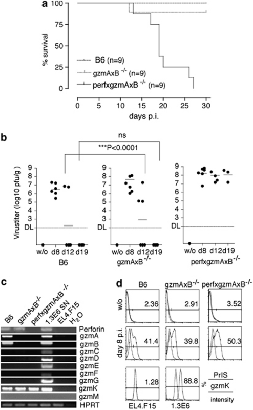
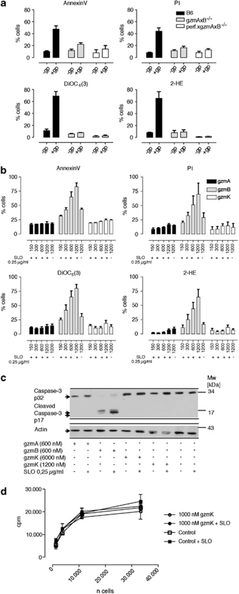
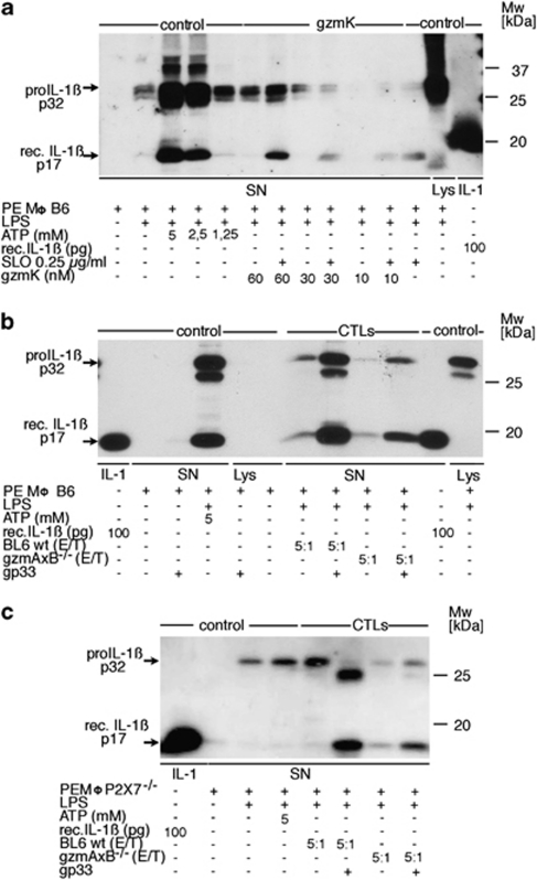
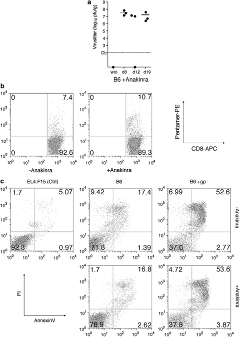
Granzymes (gzms) are key components of T-killer (Tc) cells believed to mediate pro-apoptotic activities. Recent evidence suggests that gzms also possess non-cytotoxic activities that contribute to host defense. In this study, we show that Tc cells from lymphocytic choriomeningitis virus (LCMV)-infected wild-type (wt) and gzm A/B-deficient mice express similar levels of gzmK protein, with both mouse strains efficiently controlling infection. GzmK, in recombinant form or secreted by ex vivo-derived LCMV-immune gzmAxB(-/-) Tc cells, lacks pro-apoptotic activity. Instead, gzmK induces primary mouse macrophages to process and secrete interleukin-1β, independent of the ATP receptor P2X(7). Together with the finding that IL-1Ra (Anakinra) treatment inhibits virus elimination but not generation of cytotoxic Tc cells in wt mice, the data suggest that Tc cells control LCMV through non-cytotoxic processes that involve gzmK.


Figures

Figure 1
Figure 1
(a) Survival of wild-type, gzmAxB−/− and perfxgzmAxB−/− mice infected with LCMV-WE. Groups (nine mice each) of B6 (dashed line) or gzmAxB−/−(dotted line) or perfgzmAxB−/− (line) mice were infected with 1 × 10⁵ p.f.u. LCMV-WE i.p., and survival was monitored for 30 days. (b) LCMV-WE replication in the liver of WT, gzmAxB−/− and perfxgzmAxB−/− mice. Groups of B6, gzmAxB−/− and perfxgzmAxB−/− (18 mice per strain) recipients were infected with 1 × 10⁵ p.f.u. LCMV-WE i.p. Six mice from each group were killed at 8, 12 and 19 days p.i. Virus titers of individual mice were determined by the plaque titer assay as described in the ‘Materials and Methods' section. (c) Expression of mRNA in ex vivo LCMV-immune CD8-enriched Tc cells from WT, gzmAxB−/− and perfxgzmAxB−/− mice. Total mRNA was isolated from ex vivo LCMV-immune CD8-enriched Tc cells (day 8 p.i.) from wild-type, gzmAxB−/− and perfxgzmAxB−/− mice, previously infected with 1 × 10⁵ p.f.u. LCMV-WE i.p. Samples were analyzed by RT-PCR for their expression of transcripts using specific primers for gzmA-G, gzmK, gzmM, Prf1 and Hprt1. (d) Intracellular expression of gzm and perf protein in ex vivo LCMV-immune CD8-enriched Tc cells (day 8 p.i.). Ex vivo LCMV-immune CD8-enriched Tc cells (day 8 p.i.) of wt, gzmAxB−/− and perfxgzmAxB−/− mice, infected with 1 × 10⁵ p.f.u. LCMV-WE i.p. were analyzed by FACS for intracellular expression of gzmK (blue line) using the respective rabbit anti-K immune serum and the control pre-immune serum (red line)
Figure 2
Figure 2
(a) Induction of pro-apoptotic markers by ex vivo LCMV-immune Tc cells from WT, gzmAxB−/− and perfxgzmAxB−/− mice. Gp-33-pulsed or untreated MEF target cells were incubated with ex vivo LCMV-immune CD8-enriched Tc cells of the indicated strain (4 h, 10 : 1 effector/target ratio). Subsequently, PS exposure on the plasma membrane (annexin-V-FITC) and PI uptake, and in parallel, ΔΨm loss (DiOC6) and ROS generation (2-HE) were analyzed by three-color flow cytometry in the cell population negative for CD8 expression as described in the ‘Materials and Methods' section. Data in the panels are represented as mean±S.E.M. of three independent experiments. (b) rec. (Mo)gzmA and rec. (Mo)gzmK are not cytotoxic: analyses of apoptotic parameters during (Mo)gzmA, (Mo)gzmB and (Mo)gzmK-induced cell death in EL4 cells. EL4 cells were incubated with the indicated amounts of gzms in the presence of a sublytic dose of SLO for 4 h, and the highest amount of gzm was also probed without SLO indicated by 1200 nM – SLO. Subsequently, PS exposure on the plasma membrane (annexin-V-FITC) and PI uptake, and in parallel, ΔΨm loss (DiOC6) and ROS generation (2-HE) were analyzed by three-color flow cytometry. (c) EL4 cells were either incubated or not incubated with the indicated amounts of rec. (Mo)gzmA, rec. (Mo)gzmB and rec. (Mo)gzmK in the presence and absence of SLO for 1 h at 37°C. Subsequently, lysates were prepared and caspase-3 and cleaved caspase-3 were analyzed by WB, with actin serving as control. (d) Survival assay of EL4 cells treated with rec. (Mo)gzmK in the presence and absence of SLO as described in the ‘Materials and Methods' section
Figure 3
Figure 3
(a) Induction of IL-1β release by rec. (Mo)gzmK. Adherent B6 PEMØs, previously sensitized with thioglycollate in vivo and challenged with LPS in vitro were incubated with the indicated amounts of gzmK in presence or absence of 0.25 μg/ml SLO, or ATP as positive control, for 3 h and SNs were subjected to WB analysis using anti-IL-1β-specific Ab. Aliquots of the following preparations were used as standards for WB analysis: SNs of ATP-sensitized pre-activated B6 PEMØs; SNs of thioglycollate-pre-sensitized and LPS-challenged B6 PEMØs. (b) Induction of IL-1β release by ex vivo CD8 cells expressing gzmK. Thioglycollate-pre-sensitized and LPS-challenged adherent B6 PEMØs were incubated with ex vivo-derived LCMV-immune Tc cells from B6 or gzmAxB ko mice at an E/T ratio of 5 : 1 in the presence or absence of the LCMV-specific peptide gp33. SN was taken after 6 h and subjected to WB analysis using anti-IL-1β-specific Ab. As control, SN from thioglycollate-pre-sensitized and LPS-challenged B6 PEMØs, and subsequently stimulated with 5 mM ATP, was analyzed accordingly. Aliquots of the following preparations were used as standards for WB analysis: rec. IL-1βs, p17; SN of ATP-sensitized pre-activated wt B6 PEMØs; SN of wt thioglycollate-pre-sensitized and LPS-activated B6 PEMØs. (c) Induction of IL-1β release by ex vivo CD8 cells expressing gzmK by targets lacking the P2X7 receptor. Experimental procedure was conducted as described in panel b, but with macrophages from P2X7−/− mice. Controls were prepared individually for this experiment with P2X7−/− macrophages (SN of ATP-sensitized pre-activated PEMØs; SN of wt thioglycollate-pre-sensitized and LPS-activated PEMØs)
Figure 4
Figure 4
(a) Anakinra inhibits recovery from LCMV infection. B6 mice received Anakinra before infection (1 × 10⁵ p.f.u. LCMV-WE i.p) and then on every third day thereafter. Three mice from each group were killed at 8, 12 and 19 days and hepatic viral replication determined. (b) Anakinra-treated mice infected with LCMV express levels of LCMV-specific Tc cells similar to untreated controls. Mice were first treated with Anakinra (200 μg) 1 day before infection and then every third day. After killing (day 8), splenic CD8 cells were isolated and virus-specific Tc cells were determined by pentamer staining. The livers from these mice were used to generate data reported in panel a. (c) Anakinra-treated and control Tc cells mediate similar apoptotic effects against EL4 cells. Gp-33-pulsed or untreated EL4 target cells were incubated with ex vivo LCMV-immune CD8-enriched Tc cells (4 h, 10 : 1). Subsequently, PS exposure on the plasma membrane (annexin-V-FITC) and PI uptake were analyzed by flow cytometry

References

    1. Barry M, Bleackley RC. Cytotoxic T lymphocytes: all roads lead to death. Nat Rev Immunol. 2002;2:401–409. - PubMed
    1. Grossman WJ, Revell PA, Lu ZH, Johnson H, Bredemeyer AJ, Ley TJ. The orphan granzymes of humans and mice. Curr Opin Immunol. 2003;15:544–552. - PubMed
    1. Kramer M, Simon MM. Are proteinases functional molecules of T cells. Immunol Today. 1987;8:140–143. - PubMed
    1. Hayes MP, Berrebi GA, Henkart PA. Induction of target cell DNA release by the cytotoxic T lymphocyte granule protease granzyme A. J Exp Med. 1989;170:933–946. - PMC - PubMed
    1. Shi L, Kraut RP, Aebersold R, Greenberg AH. A natural killer cell granule protein that induces DNA fragmentation and apoptosis. J Exp Med. 1992;175:553–566. - PMC - PubMed

Publication types

MeSH terms