. 2010 Dec;14(6):427-33.
doi: 10.4196/kjpp.2010.14.6.427. Epub 2010 Dec 31.

Effect of Extremely Low Frequency Electromagnetic Fields (EMF) on Phospholipase Activity in the Cultured Cells

Affiliations

Effect of Extremely Low Frequency Electromagnetic Fields (EMF) on Phospholipase Activity in the Cultured Cells

Ho Sun Song et al. Korean J Physiol Pharmacol. 2010 Dec.

Abstract

This study was conducted to investigate the effects of extremely low frequency electromagnetic fields (EMF) on signal pathway in plasma membrane of cultured cells (RAW 264.7 cells and RBL 2H3 cells), by measuring the activity of phospholipase A(2) (PLA(2)), phospholipase C (PLC) and phospholipase D (PLD). The cells were exposed to the EMF (60 Hz, 0.1 or 1 mT) for 4 or 16 h. The basal and 0.5 µM melittin-induced arachidonic acid release was not affected by EMF in both cells. In cell-free PLA(2) assay, we failed to observe the change of cPLA(2) and sPLA(2) activity. Also both PLC and PLD activities did not show any change in the two cell lines exposed to EMF. This study suggests that the exposure condition of EMF (60 Hz, 0.1 or 1 mT) which is 2.4 fold higher than the limit of occupational exposure does not induce phospholipases-associated signal pathway in RAW 264.7 cells and RBL 2H3 cells.

Keywords: Arachidonic acid; EMF; Phospholipase A2; Phospholipase C; Phospholipase D.


Figures
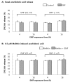
Fig. 1
Fig. 1
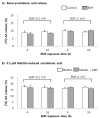
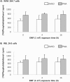
The changes of basal (A) and 0.5 µM melittin-induced [3H]AA release (B) in RAW 264.7 cells. The cells were exposed to EMF (60 Hz, 0.1 or 1 mT) for 4 or 16 h and were labeled with [3H]AA for 2 h. The radioactivity of released [3H]AA was measured in the presence or absence of 0.5 µM melittin. Results are indicated in mean±S.D. from four separate experiments.
Fig. 2
Fig. 2
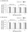
The changes of basal (A) and 0.5 µM melittin-induced [3H]AA release (B) in RBL 2H3 cells. The cells were exposed to EMF (60 Hz, 0.1 or 1 mT) for 4 or 16 h and were labeled with [3H]AA for 2 h. The radioactivity of released [3H]AA was measured in the presence or absence of 0.5 µM melittin. Results indicate mean±S.D. from four separate experiments.
Fig. 3
Fig. 3
Cell-derived PLA2 activity in the presence of 5 mM CaCl2. PLA2 (25 µg protein) derived from RAW 264.7 cells and RBL 2H3 cells was incubated with 1-palmitoyl-2-[14C]arachidonyl phosphatidylcholine in the presence of 10 µM AACOCF3 (cPLA2 inhibitor) or 1 mM DTT (sPLA2 inhibitor) and in the absence of CaCl2. Results indicate mean±S.D. from four separate experiments. *Significantly different from Control (p<0.05).
Fig. 4
Fig. 4
Effect of EMF on cell-derived cPLA2 activity. Cell-derived cPLA2 was obtained from the RAW 264.7 cells (A) and RBL 2H3 cells (B) exposed to EMF (60 Hz, 0.1 or 1 mT) for 4 or 16 h. cPLA2 (25 µg protein) was incubated with 1-palmitoyl-2-[14C]arachidonyl phosphatidylcholine in the presence of 5 mM CaCl2 and 1 mM DTT. Results indicate mean±S.D. from four separate experiments.
Fig. 5
Fig. 5
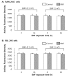
Effect of EMF on cell-derived sPLA2 activity. Cell-derived sPLA2 was obtained from the RAW 264.7 cells (A) and RBL 2H3 cells (B) exposed to EMF (60 Hz, 0.1 or 1 mT) for 4 or 16 h. sPLA2 (100 µg protein) was incubated with 10-pyren phosphatidylcholine. Results indicate mean±S.D. from four separate experiments.
Fig. 6
Fig. 6
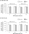
Effect of EMF on cell-derived PLC activity. Cell-derived PLC was obtained from the RAW 264.7 cells (A) and RBL 2H3 cells (B) exposed to EMF (60 Hz, 0.1 or 1 mT) for 4 or 16 h. PLC (25 µg protein) was incubated with [3H]phosphatidylinositol. Results indicate mean±S.D. from four separate experiments.
Fig. 7
Fig. 7
Effect of EMF on PLD activity. RAW 264.7 cells (A) and RBL 2H3 cells (B) were exposed to EMF (60 Hz, 1 mT) for 4 or 16 hand were labeled with [3H]oleic acid, for 3 h. The radioactivity of [3H]phosphatidylethanol produced by PLD was measured in the presence or absence of 1 µM PMA. Results indicate mean±S.D. from four separate experiment.

References

    1. Wertheimer N, Leeper E. Electrical wiring configurations and childhood cancer. Am J Epidemiol. 1979;109:273–284. - PubMed
    1. Floderus B, Persson T, Stenlund C, Wennberg A, Ost A, Knave B. Occupational exposure to electromagnetic fields in relation to leukemia and brain tumors: a case-control study in Sweden. Cancer Causes Control. 1993;4:465–476. - PubMed
    1. Matanoski GM, Elliott EA, Breysse PN, Lynberg MC. Leukemia in telephone linemen. Am J Epidemiol. 1993;137:609–619. - PubMed
    1. Glaser KB. Regulation of phospholipase A2 enzymes: selective inhibitors and their pharmacological potential. Adv Pharmacol. 1995;32:31–66. - PubMed
    1. Attur MG, Patel R, Thakker G, Vyas P, Levartovsky D, Patel P, Naqvi S, Raza R, Patel K, Abramson D, Bruno G, Abramson SB, Amin AR. Differential anti-inflammatory effects of immunosuppressive drugs: cyclosporin, rapamycin and FK-506 on inducible nitric oxide synthase, nitric oxide, cyclooxygenase-2 and PGE2 production. Inflamm Res. 2000;49:20–26. - PubMed