. 2011 Aug;19(8):1239-42.
doi: 10.1007/s00167-011-1426-z. Epub 2011 Feb 11.

Advances in the three-portal technique for anatomical single- or double-bundle ACL reconstruction

Affiliations

Advances in the three-portal technique for anatomical single- or double-bundle ACL reconstruction

Paulo H Araujo et al. Knee Surg Sports Traumatol Arthrosc. 2011 Aug.

Abstract

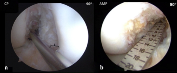
Purpose: To describe the "three-portal technique for anatomical ACL single- or double-bundle reconstruction" and the arthroscopic viewing improvement provided by this technique.

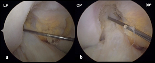
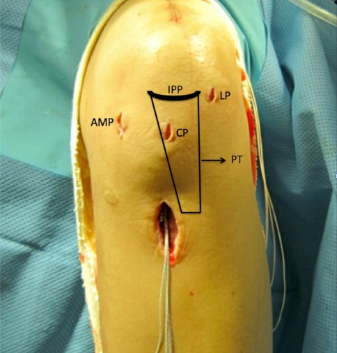
Methods: A "high" anterolateral portal was placed 1 cm lateral to the patellar tendon and the most inferior portion of the portal at the level of the inferior pole of the patella. A "central" portal was placed using a spinal needle under arthroscopic visualization following the orientation of the previous ACL fibers. An accessory medial portal was also placed using a spinal needle respecting a 2-mm distance to the medial femoral condyle.

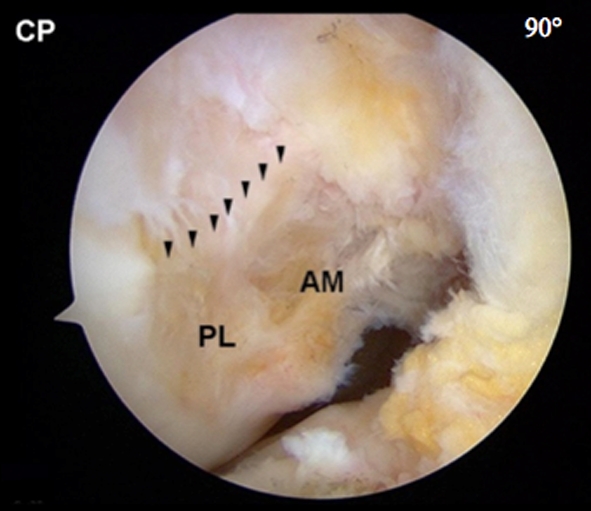
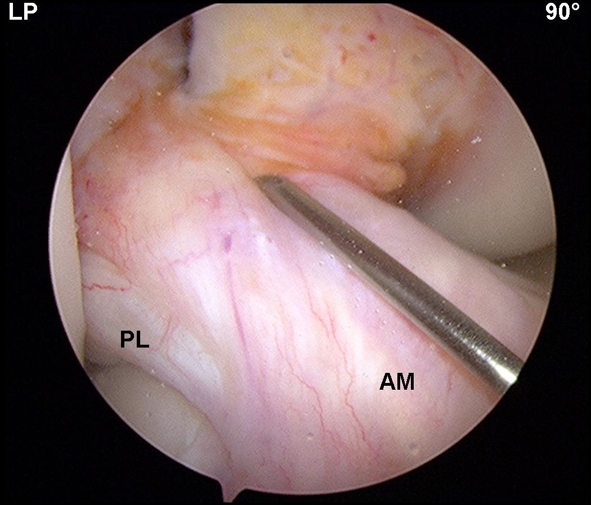
Results: The "high" anterolateral portal permitted a broad and unobstructed view of the ACL tibial attachment. The "central" portal allowed a straightforward view of the ACL femoral remnant and bony landmarks in the intercondylar notch. The accessory medial portal enabled to reach the femoral native insertion site of the ACL.

Conclusion: The three-portal technique provides a proper view of the soft tissue remnants and bony landmarks facilitating an anatomical positioning of the graft.


Figures

Fig. 1
Fig. 1
Arthroscopic view of the right knee in 90° of flexion. a Anterolateral portal view (LP) is unable to visualize the femoral ACL insertion site. b Central anteromedial portal view (CP) provides adequate visualization and shows the guide pin is located outside of the native ACL insertion site
Fig. 2
Fig. 2
Left knee in operating position. The three portals and their localization according to the inferior pole of the patella (IPP) and patellar tendon (PT). LP high anterolateral portal, CP central anteromedial portal, AMP accessory anteromedial portal
Fig. 3
Fig. 3
a “High” anterolateral portal positioning. b Arthroscopic view of the tibial ACL insertion site of the right knee from the high anterolateral portal. IPP  inferior pole of the patella, LP high anterolateral portal, AM tibial anteromedial bundle insertion site, PL tibial posterolateral bundle insertion site
Fig. 4
Fig. 4
Central anteromedial portal (CP) view of the femoral insertion site of the right knee. The lateral intercondylar ridge can be visualized, as well as the anteromedial (AM) and posterolateral (PL) bundle insertion sites
Fig. 5
Fig. 5
The central anteromedial portal (CP) is created under direct visualization through the anterolateral (LP) portal and follows the ACL fibers orientation. AM anteromedial, PL posterolateral bundle
Fig. 6
Fig. 6
a Central anteromedial portal (CP) view of the femoral ACL insertion site of the right knee. The guide pin is in the accessory anteromedial portal. There should be at least 2 mm between the guide pin and the medial femoral condyle (bracket) to ensure safe femoral tunnel drilling without damaging the cartilage. b Under direct visualization through the accessory medial portal, the ruler is inserted through the high anterolateral portal (LP) to measure the femoral ACL insertion site size. CP central anteromedial portal view, AMP accessory medial portal view

References

    1. Schreiber VM, van Eck CF, Fu FH. Anatomic double-bundle ACL reconstruction. Sports Med Arthrosc Rev. 2010;18:27–32. doi: 10.1097/JSA.0b013e3181bf6634. - DOI - PubMed
    1. Bedi A, Altchek DW. The “Footprint” anterior cruciate ligament technique: an anatomic approach to anterior cruciate ligament reconstruction. Arthroscopy. 2009;25:1128–1138. doi: 10.1016/j.arthro.2009.03.008. - DOI - PubMed
    1. Lohmander LS, Ostenberg A, Englund M, Roos H. High prevalence of knee osteoarthritis, pain, and functional limitations in female soccer players twelve years after anterior cruciate ligament injury. Arthritis Rheum. 2004;50:3145–3152. doi: 10.1002/art.20589. - DOI - PubMed
    1. Nikolaou VS, Efstathopoulos N, Sourlas I, Pilichou A, Papachristou G. Anatomic double-bundle versus single-bundle ACL reconstruction: a comparative biomechanical study in rabbits. Knee Surg Sports Traumatol Arthrosc. 2009;17:895–906. doi: 10.1007/s00167-009-0754-8. - DOI - PubMed
    1. Snow M, Stanish WD. Double-bundle ACL reconstruction: how big is the learning curve? Knee Surg Sports Traumatol Arthrosc. 2010;18:1195–1200. doi: 10.1007/s00167-010-1062-z. - DOI - PubMed

MeSH terms