. 2011 Mar;41(3):863-72.
doi: 10.1002/eji.201040649. Epub 2011 Feb 11.

Interferon-α induces unabated production of short-lived plasma cells in pre-autoimmune lupus-prone (NZB×NZW)F1 mice but not in BALB/c mice

Affiliations

Interferon-α induces unabated production of short-lived plasma cells in pre-autoimmune lupus-prone (NZB×NZW)F1 mice but not in BALB/c mice

Alexis Mathian et al. Eur J Immunol. 2011 Mar.

Abstract

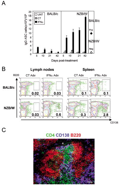
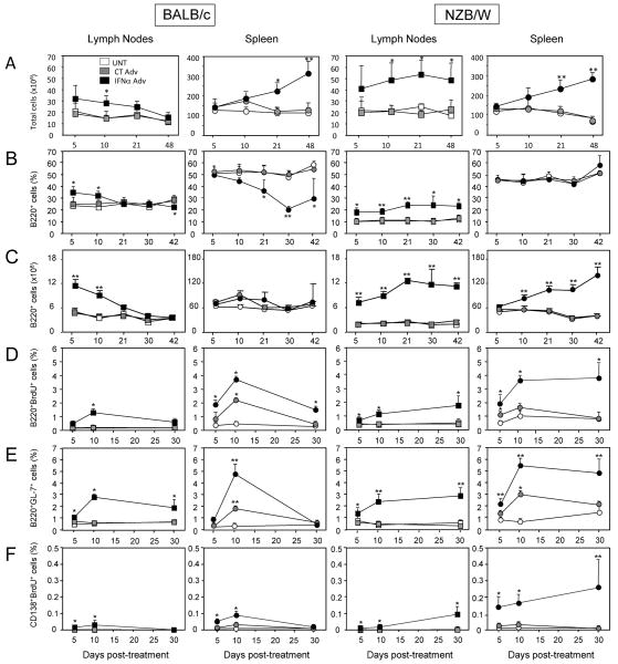
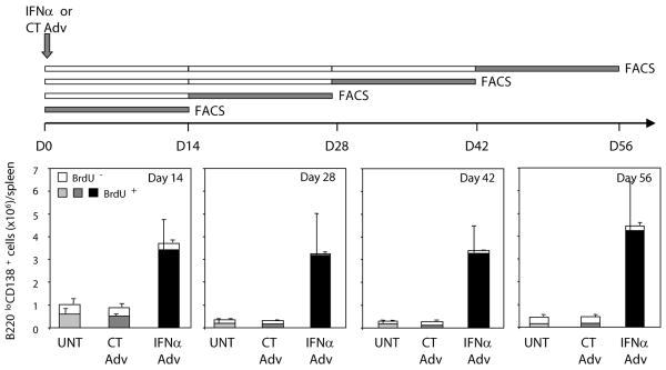
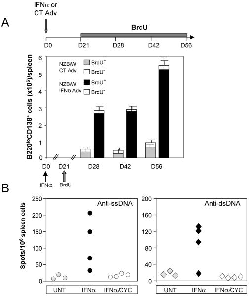
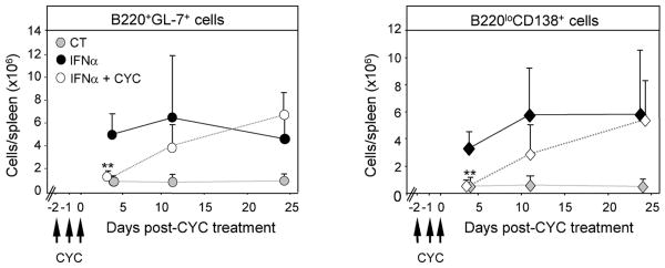
IFN-α is known to play a critical role in the pathogenesis of systemic lupus erythematosus (SLE), but the mechanisms remain unclear. We previously showed that within weeks, exposure to IFN-α in vivo induces lupus in pre-autoimmune lupus-prone NZB×NZW F1 (NZB/W) but not in BALB/c mice. In the current study, we show that in vivo expression of IFN-α induces sustained B-cell proliferation in both BALB/c and NZB/W mice. In NZB/W but not BALB/c mice, B-cell proliferation was accompanied by a rapid and unabated production of autoantibody-secreting cells (ASCs) in secondary lymphoid organs, suggesting that a B-cell checkpoint is altered in the autoimmune background. The majority (>95%) of ASCs elicited in IFN-α-treated NZB/W mice were short-lived and occurred without the induction of long-lived plasma cells. A short course of cyclophosphamide caused a sharp drop in IFN-α-elicited short-lived plasma cells, but the levels recovered within days following termination of treatment. Thus, our work provides new insights into effectiveness and limitations of the current SLE therapies.


Conflict of interest statement

Conflicts of interest: The authors declare no conflict of interest.

Figures

Figure 1
Figure 1. IFNα in vivo elicits long-term production of ASCs in NZB/W but not in BALB/c mice
(A) Frequency of spleen IgG ASCs was determined by ELISPOT. Results are the means ± SD of three mice in each group from two independent experiments. Neg, young untreated NZB/W mice; pos, old proteinuric untreated NZB/W mice; UNT, untreated; CT, control adenovirus (CT Adv)-treated; IFNα, interferon alpha (IFNα Adv)-treated. (B) Spleen and lymph node cells were stained with fluorescent anti-B220 and anti-CD138 Abs, and the frequency of ASCs analyzed by FACS at day 42 post-treatment. Data shown are representative of eight experiments (C) Immunofluorescence staining of plasma cells in spleen sections from IFNα-treated NZB/W mice for CD4 (green), CD138 (blue), and B220 (red). T, T cell zone; J, junction of the T cell zone; R, red pulp; G, germinal center. Data shown are representative of four experiments.
Figure 2
Figure 2. IFNα elicits sustained B lymphocyte and plasmablast proliferation as well as GC formation in young NZB/W mice but not in Balb/c mice
(A) White cell counts in secondary lymphoid organ in Balb/c and NZB/W mice. (B) Frequency and (C) absolute number of B220+CD19+ B cells in the spleen and lymph nodes of Balb/c and NZB/W mice as determined by FACS. (D) Proliferation of B cells was assessed by FACS of B220+BrdU+ cells from mice injected with BrdU 2-3 h before sacrifice at the indicated times. (E) Frequency of GC B cells was determined by FACS of B220+GL-7+ cells. (F) Frequency of proliferating plasmablasts was determined by FACS of B220lowCD138+BrdU+ cells from mice injected with BrdU 2 to 3 h before sacrifice at the indicated times. Results are means ± SD of 3 to 5 mice/experimental group from 2 independent experiments and are expressed as % of total cells analyzed. Comparisons between IFNα Adv-treated and CT Adv-treated animals and between CT Adv-treated and untreated animals were made using the Mann-Whitney U test. *, p<0.05; **, p<0.01.
Figure 3
Figure 3. IFNα in vivo promotes the continuous generation of ASCs
Four groups each of untreated (UNT), CT Adv-treated (CT), or IFNα Adv-treated (IFNα) NZB/W mice (at day 0) were fed BrdU for different 14-day periods (days 0-14, 14-28, 28-42, and 56-70) at the end of which mice were sacrificed and spleen cells were stained for intracellular BrdU (top panel), and BrdU staining of gated B220loCD138+ cells were analyzed by FACS. Raw results (see Supplemental Figure 6) were converted into absolute numbers of BrdU-negative (white columns) and BrdU-positive (shaded columns) B220loCD138+ cells/spleen and are the means ± SD of 3 to 4 mice per group based on three independent experiments.
Figure 4
Figure 4. IFNα treatment elicits short-lived ASCs in NZB/W mice
(A) Absence of long-lived plasma cells. NZB/W mice were treated at day 0 with CT or IFNα Adv and then continuously fed BrdU for 35 days starting at day 21 post-treatment. Mice were sacrificed at indicated times, and BrdU staining of B220lowCD138+ cells was analyzed by FACS. Results are means ± SD of 3 to 5 mice per experimental group from two independent experiments (B) NZB/W mice were untreated (UNT) or treated with IFNα Adv and either with PBS (IFNα) or with 20 mg/kg cyclophosphamide (IFNα/CYC) on 3 consecutive days starting on day 21 after initiation of IFNα treatment. Mice were sacrificed 4 days after the last injection of cyclophosphamide, and spleen anti-ssDNA (left) or anti-dsDNA(right) ASCs were measured by ELISPOT. Each symbol represents an individual mouse. Data shown are representative of three independent experiments.
Figure 5
Figure 5. Effect of a short course of cyclophosphamide on IFNα-induced B cell proliferation and ASC production
IFNα Adv-treated NZB/W mice were injected with 20 mg/kg cyclophosphamide (IFNα+CYC) for 3 consecutive days starting at 21 days after initiation of IFNα treatment. Controls consisted of CT Adv-treated NZB/W mice (CT) and IFNα-Adv-treated NZB/W mice that had not received cyclophosphamide (IFNα). At indicated times, the numbers of spleen B220+GL-7+ GC B cells and B220loCD138+ ASCs were determined by FACS. Results are the means ± SD of 3 to 5 mice per group based on three independent experiments. Comparisons between IFNα Adv-treated and IFNα Adv + CYC-treated animals were made using the Mann-Whitney U test. **, p<0.01.

Comment in

References

    1. Hahn BH. Antibodies to DNA. N Engl J Med. 1998;338:1359–1368. - PubMed
    1. Ronnblom L, Eloranta ML, Alm GV. The type I interferon system in systemic lupus erythematosus. Arthritis Rheum. 2006;54:408–420. - PubMed
    1. Braun D, Geraldes P, Demengeot J. Type I Interferon controls the onset and severity of autoimmune manifestations in lpr mice. J Autoimmun. 2003;20:15–25. - PubMed
    1. Santiago-Raber ML, Baccala R, Haraldsson KM, Choubey D, Stewart TA, Kono DH, Theofilopoulos AN. Type-I interferon receptor deficiency reduces lupus-like disease in NZB mice. J Exp Med. 2003;197:777–788. - PMC - PubMed
    1. Mathian A, Weinberg A, Gallegos M, Banchereau J, Koutouzov S. IFN-alpha induces early lethal lupus in preautoimmune (New Zealand Black × New Zealand White) F1 but not in BALB/c mice. J Immunol. 2005;174:2499–2506. - PubMed

Publication types

MeSH terms