

Substantia nigra hyperechogenicity with LRRK2 G2019S mutations
- PMID: 21312285
- PMCID: PMC3082617
- DOI: 10.1002/mds.23644
Substantia nigra hyperechogenicity with LRRK2 G2019S mutations
Abstract
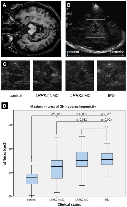
Background: Transcranial sonography (TCS) area of hyperechogenicity in the substantia nigra (aSN) is increased in idiopathic and genetic Parkinson's disease (PD).
Methods: We performed TCS in 34 LRRK2 G2019S mutation carriers manifesting PD, 24 non-manifesting mutation carriers, and 28 idiopathic PD patients and compared them with 40 healthy controls (total, n = 126).
Results: Compared with the controls (mean 0.15 cm(2) ), the aSN values in all other groups were increased. The mean aSN was 0.23 cm(2) in nonmanifesting mutation carriers (P = .015), 0.34 cm(2) in idiopathic PD patients (P < .0001), 0.32 cm(2) in LRRK2-associated PD patients (P < .0001), and 0.33 cm(2) in the overall PD group (P < .0001). LRRK2-associated PD patients had a higher aSN than did nonmanifesting carriers (P = .011), but there was no significant difference in aSN between patients with idiopathic and LRRK2-associated PD (P = .439).
Conclusions: Our results suggest that SN pathoanatomical alterations may not be substantially different between idiopathic and LRRK2-associated PD. The findings in the nonmanifesting mutation carriers suggest the presence of intermediate nigrostriatal pathology consistent with the age-dependent reduced penetrance of this mutation.
Copyright © 2011 Movement Disorder Society.
Figures
References
-
- Walter U, Behnke S, Eyding J, et al. Transcranial brain parenchyma sonography in movement disorders: state of the art. Ultrasound Med Biol. 2007;33(1):15–25. - PubMed
-
- Hagenah JM, Becker B, Brüggemann N, et al. Transcranial sonography findings in a large family with homozygous and heterozygous PINK1 mutations. J Neurol Neurosurg Psychiatry. 2008;79(9):1071–1074. - PubMed
-
- Hagenah JM, Konig IR, Becker B, et al. Substantia nigra hyperechogenicity correlates with clinical status and number of Parkin mutated alleles. J Neurol. 2007;254(10):1407–1413. - PubMed
-
- Schweitzer KJ, Brussel T, Leitner P, et al. Transcranial ultrasound in different monogenetic subtypes of Parkinson’s disease. J Neurol. 2007;254(5):613–616. - PubMed
-
- Walter U, Klein C, Hilker R, Benecke R, Pramstaller PP, Dressler D. Brain parenchyma sonography detects preclinical parkinsonism. Mov Disord. 2004;19(12):1445–1449. - PubMed
Publication types
MeSH terms
Substances
Grants and funding
LinkOut - more resources
Full Text Sources
Medical
