

Formulation and characterisation of magnetic resonance imageable thermally sensitive liposomes for use with magnetic resonance-guided high intensity focused ultrasound
- PMID: 21314334
- PMCID: PMC3417228
- DOI: 10.3109/02656736.2010.528140
Formulation and characterisation of magnetic resonance imageable thermally sensitive liposomes for use with magnetic resonance-guided high intensity focused ultrasound
Abstract
Purpose: Objectives of this study were to: 1) develop iLTSL, a low temperature sensitive liposome co-loaded with an MRI contrast agent (ProHance® Gd-HP-DO3A) and doxorubicin, 2) characterise doxorubicin and Gd-HP-DO3A release from iLTSL and 3) investigate the ability of magnetic resonance-guided high intensity focused ultrasound (MR-HIFU) to induce and monitor iLTSL content release in phantoms and in vivo.
Methods: iLTSL was passively loaded with Gd-HP-DO3A and actively loaded with doxorubicin. Doxorubicin and Gd-HP-DO3A release was quantified by fluorescence and spectroscopic techniques, respectively. Release with MR-HIFU was examined in tissue-mimicking phantoms containing iLTSL and in a VX2 rabbit tumour model.
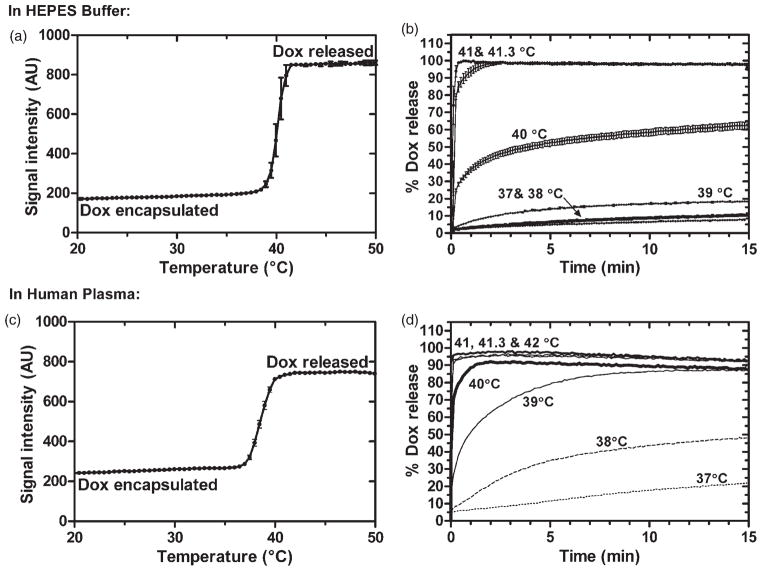
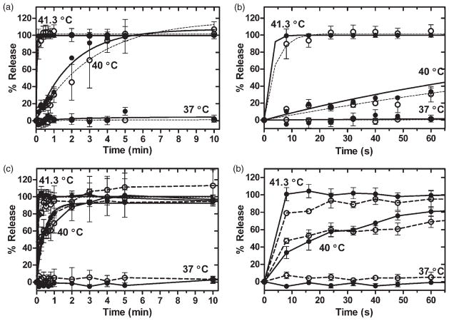
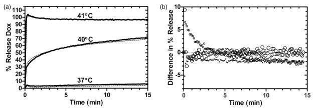
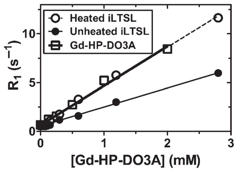
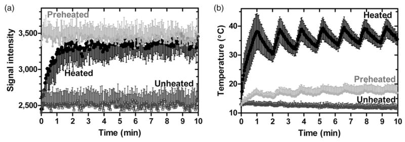
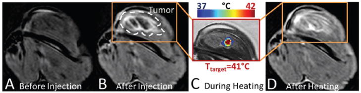
Results: iLTSL demonstrated consistent size and doxorubicin release kinetics after storage at 4°C for 7 days. Release of doxorubicin and Gd-HP-DO3A from iLTSL was minimal at 37°C but fast when heated to 41.3°C. The magnitude of release was not significantly different between doxorubicin and Gd-HP-DO3A over 10 min in HEPES buffer and plasma at 37°, 40° and 41.3°C (p > 0.05). Relaxivity of iLTSL increased significantly (p < 0.0001) from 1.95 ± 0.05 to 4.01 ± 0.1 mMs⁻¹ when heated above the transition temperature. Signal increase corresponded spatially and temporally to MR-HIFU-heated locations in phantoms. Signal increase was also observed in vivo after iLTSL injection and after each 10-min heating (41°C), with greatest increase in the heated tumour region.
Conclusion: An MR imageable liposome formulation co-loaded with doxorubicin and an MR contrast agent was developed. Stability, imageability, and MR-HIFU monitoring and control of content release suggest that MR-HIFU combined with iLTSL may enable real-time monitoring and spatial control of content release.
Conflict of interest statement
NIH may have intellectual property ion the field. NIH and Philips Healthcare have a cooperative research and development agreement. AP is a salaried employee of Philips Healthcare.
Figures
References
-
- Allen TM, Cullis PR. Drug delivery systems: Entering the mainstream. Science. 2004;303:1818–1822. - PubMed
-
- Tilcock C. Delivery of contrast agents for magnetic resonance imaging, computed tomography, nuclear medicine and ultrasound. Adv Drug Deliv Rev. 1999;37:33–51. - PubMed
-
- Maeda H, Seymour LW, Miyamoto Y. Conjugates of anticancer agents and polymers - advantages of macromolecular therapeutics in vivo. Bioconjugate Chem. 1992;3:351–362. - PubMed
-
- Matsumura Y, Maeda H. A new concept for macromolecular therapeutics in cancer-chemotherapy - mechanism of tumori-tropic accumulation of proteins and the antitumor agent SMANCS. Cancer Res. 1986;46:6387–6392. - PubMed
-
- Northfelt DW, Martin FJ, Working P, Volberding PA, Russell J, Newman M, Amantea MA, Kaplan LD. Doxorubicin encapsulated in liposomes containing surface-bound polyethylene glycol: Pharmacokinetics, tumor localization, and safety in patients with AIDS-related Kaposi’s sarcoma. J Clin Pharmacol. 1996;36:55–63. - PubMed
Publication types
MeSH terms
Substances
Grants and funding
LinkOut - more resources
Full Text Sources
Other Literature Sources
Research Materials
Miscellaneous