Review
. 2011:40:169-86.
doi: 10.1146/annurev-biophys-042910-155359.

Actin structure and function

Affiliations
Review

Actin structure and function

Roberto Dominguez et al. Annu Rev Biophys. 2011.

Abstract

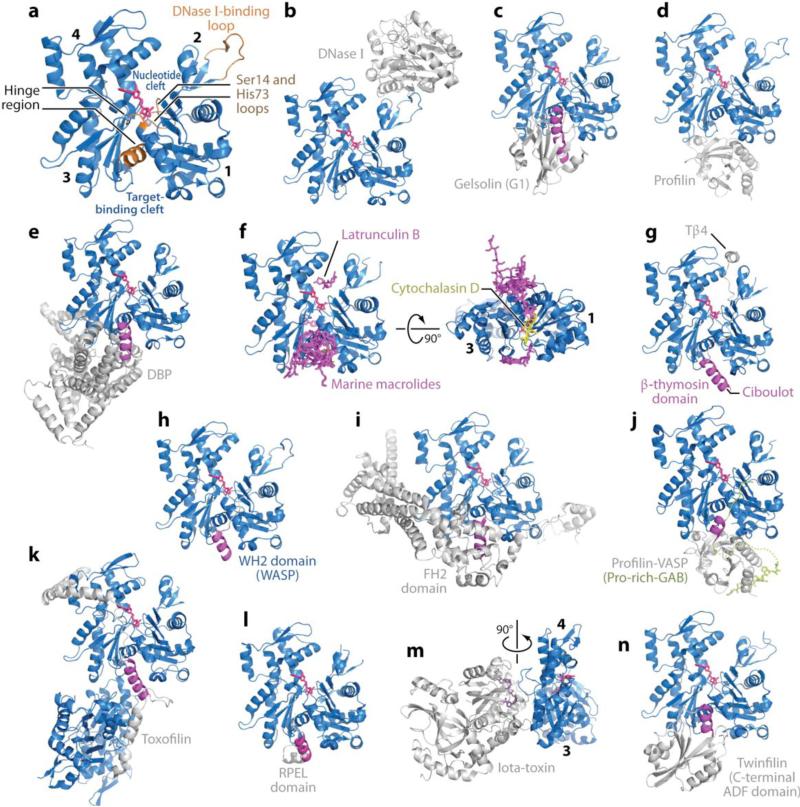
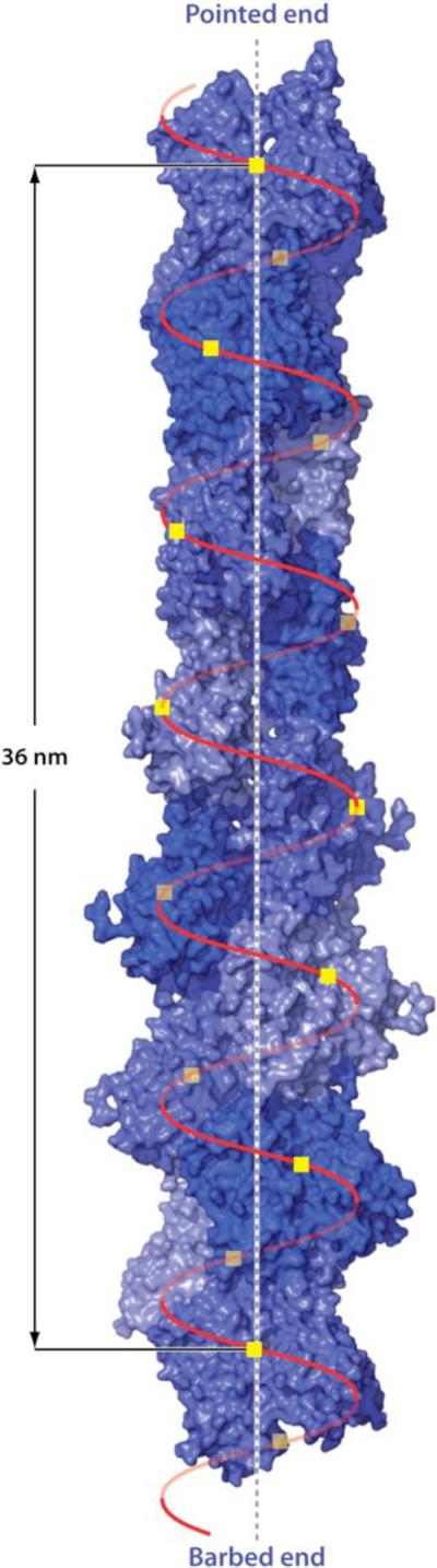
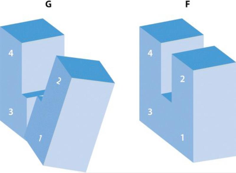
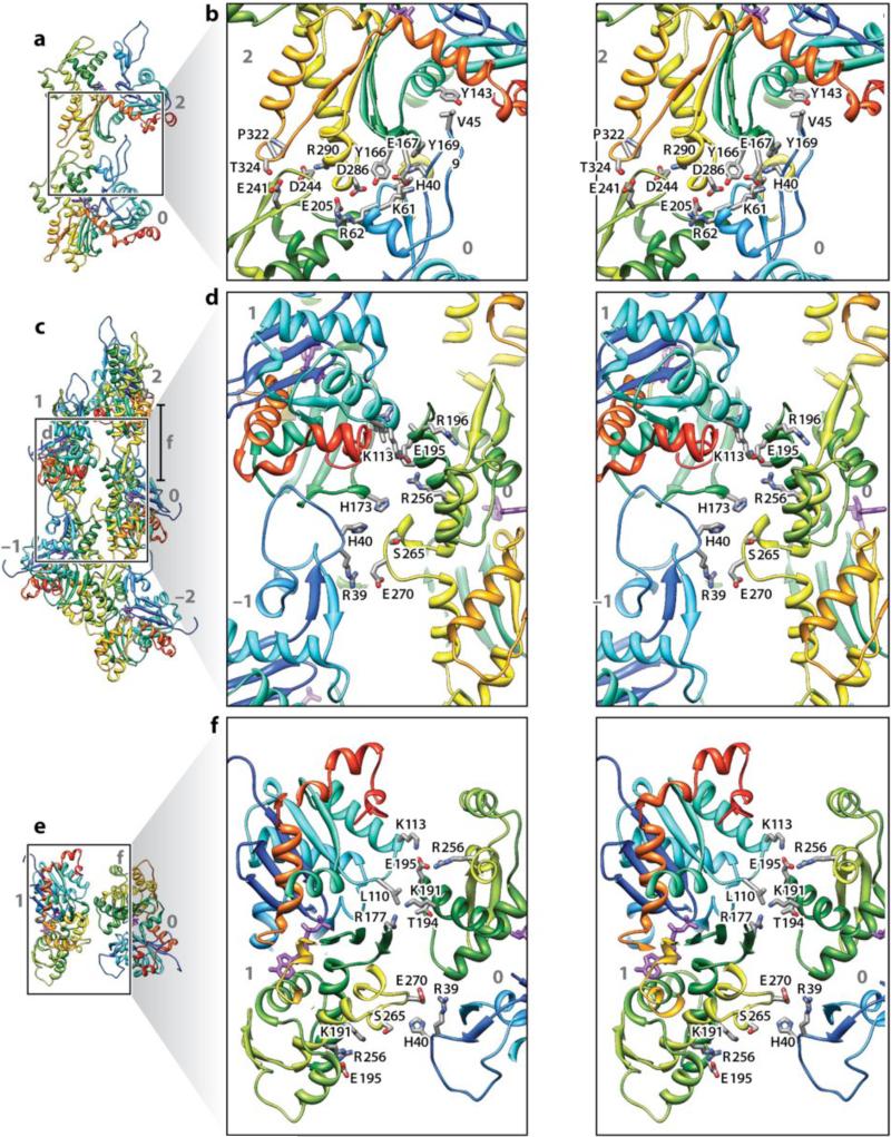
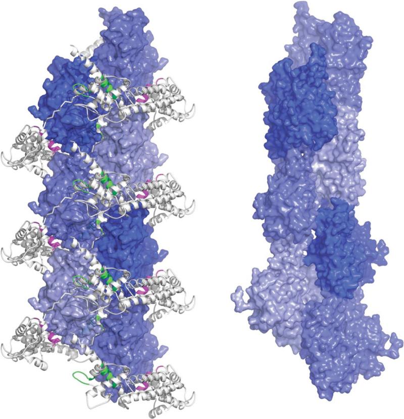
Actin is the most abundant protein in most eukaryotic cells. It is highly conserved and participates in more protein-protein interactions than any known protein. These properties, along with its ability to transition between monomeric (G-actin) and filamentous (F-actin) states under the control of nucleotide hydrolysis, ions, and a large number of actin-binding proteins, make actin a critical player in many cellular functions, ranging from cell motility and the maintenance of cell shape and polarity to the regulation of transcription. Moreover, the interaction of filamentous actin with myosin forms the basis of muscle contraction. Owing to its central role in the cell, the actin cytoskeleton is also disrupted or taken over by numerous pathogens. Here we review structures of G-actin and F-actin and discuss some of the interactions that control the polymerization and disassembly of actin.


Figures

Figure 1
Figure 1
Structures of actin and actin complexes. The structures of actin complexes are shown to scale and in chronological order of publication. (a) Classical view of the structure of the actin monomer. The structure shown was derived from the complex with DNase I, with completion of the C terminus from the complex of actin with profilin. Highlighted in orange are the Ser14 and methylated His73 loops, the DNase I-binding loop, and the hinge between domains, consisting of helix Gln137-Ser145 and the loop centered at residue Lys336. Subdomains 1--4 are labeled. (They are also labeled in panels f and m, which show rotated views of the structure.) Together, subdomains 1 and 2 form the outer (or small) domain, whereas subdomains 3 and 4 constitute the inner (or large) domain. Two large clefts are formed between these domains: the nucleotide- and target-binding clefts. Most actin-binding proteins (ABPs) and small molecules bind in the target-binding cleft, and the interaction frequently involves an α-helix (magenta). (b) DNase I (1ATN). (c) Gelsolin segment 1 (G1) (1EQY). See also Supplemental Figure 1 for structures of actin with Gelsolin fragments G1--G3 and G4--G6. (d) β-actin-profilin (2BTF). (e) Vitamin D-binding protein ( DBP ) (1KXP). (f) Two perpendicular views of a superimposition of structures of actin complexes with small molecules, including marine toxins (1QZ5, 1QZ5, 1S22, 1YXQ, 2ASM, 2ASO, 2ASP, 2FXU, 2Q0R, 2Q0U, 2VYP), Latrunculin B [DOUG: Add “A/B’ – missing.] (2Q0U) and cytochalasin D (3EKS). The marine toxins (magenta) bind at the ends of the target-binding cleft, whereas cytochalasin D binds in the middle, and Latrunculin (both A and B) binds in the nucleotide cleft. All these molecules compromise actin polymerization. (g) β-thymosin domain. A complete structure of this complex is not available, but combined, the structures of the N-terminal portion of a β-thymosin domain from Drosophila ciboulot (1SQK) and the C-terminal end of b-thymosin peptide (Tβ4) (1T44) provide a model of this complex. (h) WASP homology domain 2 (WH2) domain of WASP (2A3Z). The WH2 domain, present in many cytoskeletal proteins in the form of tandem repeats, is related to the β-thymosin domain but lacks the C-terminal pointed end capping helix. (i) Formin homology 2 (FH2) domain (1Y64). See also Supplemental Figure 2 for a more detailed representation of this structure. (j) Ternary complex with profilin and the Pro-rich G-actin-binding (Pro-rich-GAB) domains of VASP (2PBD). The GAB domain is related to the WH2 domain but presents a shorter N-terminal helix and adopts a slightly different orientation when bound to actin, possibly because it is designed to co-bind with profilin. (k) Toxofilin from Toxoplasma gondii bound to an antiparallel actin dimer (2Q97). (l) RPEL (RPxxxEL-containing motif)[AU: Does this need to be spelled out for the general reader?] domain from the serum response factor coactivator MAL (2V52). (m) Arginine ADP-ribosylation iota-toxin from Clostridium perfringens (3BUZ). View rotated 90° relative to the other complexes. (n) C-terminal ADF/cofilin domain of twinfilin (3DAW). See Supplemental Table 1 for a complete list of references.
Figure 2
Figure 2
The helical structure of F-actin derived from cryo-electron microscopy (16). The molecules are arranged on a single helix with 13 molecules repeating in almost exactly six left-handed turns. The rise per molecule is 2.76 nm and the twist per molecule is –166.6±0.6°(for simplicity of drawing, in the figure the value -166.15 has been used to make the structure repeat exactly after 13 residues). Because -166° is close to 180° the structure takes on the appearance of a two-start right-handed long-pitch helix.
Figure 3
Figure 3
The essence of the G- to F-actin transition is a flattening of the actin molecule by a propeller-like twist of the outer and inner domains about an axis roughly at right angles to the actin helix axis. The numbers refer to the subdomains (28) (diagram courtesy of Y. Maeda).
Figure 4
Figure 4
Intermolecule bonding in F-actin. Shown are five actin molecules labeled –2 to +2. The run of the protein chain is shown as a secondary structure cartoon color-coded from blue (N terminus) to red (C terminus). Interacting side chains are shown as sticks. Panels b, d, and f are stereo pairs. Panels a and b show the main longitudinal interface between molecules 0 and 2. Panels c and d show the transverse interaction across the helix axis in the neighborhood of the plug. This interaction also has contributions from the D-loop. Panels e and f show the transverse interaction higher up (see right hand side of panel c for a definition of panel f) involving loops 195--198 and 108--113. (Reprinted by permission from Macmillan Publishers Ltd: Nature 467:724-728, copywrite 2010)
Figure 5
Figure 5
Precursor helix assembled by the formin FH2 domain. The left view shows the precursor helix assembled by FH2 subunits along the crystallographic C2 axis. The forming FH2 domain is shown grey as in Figure 1i. Neighboring molecules are positioned by binding respectively to the knob (magenta)[ and the post (green) to form a helix with a twist of 180° per molecule and a rise per residue of 28.1 Å (see Supplemental Figure 2 for details). The right view shows the F-actin helix for comparison. Actin subunits of the filament helix are rotated by -166.6° and translated by 27.6 Å (as in Figure 2)

References

    1. Barth H, Stiles BG. Binary actin-ADP-ribosylating toxins and their use as molecular Trojan horses for drug delivery into eukaryotic cells. Curr. Med. Chem. 2008;15:459–-69. - PubMed
    1. Bernstein BW, Bamburg JR. ADF/cofilin: a functional node in cell biology. Trends Cell Biol. 2010;20:187–-95. - PMC - PubMed
    1. Bork P, Sander C, Valencia A. An ATPase domain common to prokaryotic cell cycle proteins, sugar kinases, actin, and hsp70 heat shock proteins. Proc. Natl. Acad. Sci. USA. 1992;89:7290–-94. - PMC - PubMed
    1. Burtnick LD, Koepf EK, Grimes J, Jones EY, Stuart DI, et al. The crystal structure of plasma gelsolin: implications for actin severing, capping, and nucleation. Cell. 1997;90:661–-70. - PubMed
    1. Campellone KG, Welch MD. A nucleator arms race: cellular control of actin assembly. Nat. Rev. Mol. Cell Biol. 2010;11:237–-51. - PMC - PubMed

Publication types

LinkOut - more resources