

The role of cell plasticity in progression and reversal of renal fibrosis
- PMID: 21314743
- PMCID: PMC3101488
- DOI: 10.1111/j.1365-2613.2011.00760.x
The role of cell plasticity in progression and reversal of renal fibrosis
Abstract
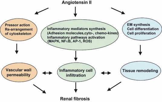
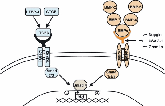
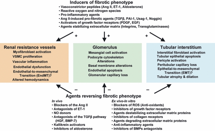
The need for novel insights into the mechanisms of progression of renal disease has become urgent during the last several years because of the increasing incidence of chronic renal disease worldwide. Independent of the underlying disease, the subsequent progression of renal fibrosis is characterized mainly by both an exaggerated synthesis and abnormal accumulation of extracellular matrix proteins produced by mesenchymal cells within the kidney. These cells are mainly myofibroblasts deriving from a variety of renal cells such as vascular smooth muscle, mesangial, resident stem, tubular epithelial, vascular endothelial cells or pericytes. The appearance of myofibroblasts is a reversible process, as suggested by studies in experimental models showing regression of renal fibrosis during therapy with antagonists and/or blockers of the renin-angiotensin system. An additional factor that can also affect the mechanisms of progression/regression of fibrosis is the plasticity of podocytes controlling glomerular filtration.
© 2011 The Authors. International Journal of Experimental Pathology © 2011 International Journal of Experimental Pathology.
Figures
References
-
- Adamczak M, Gross ML, Krtil J, et al. Reversal of glomerulosclerosis after high-dose enalapril treatment in subtotally nephrectomized rats. J. Am. Soc. Nephrol. 2003;14:2833–2842. - PubMed
-
- Anonymous. Incidence and prevalence of ESRD – United States Renal Data System. Am. J. Kidney Dis. 1998;32(Suppl. 1):S38–S49. - PubMed
-
- Arciniegas E, Sutton AB, Allen TD, Schor AM. Transforming growth factor beta 1 promotes the differentiation of endothelial cells into smooth muscle-like cells in vitro. J. Cell Sci. 1992;103:521–529. - PubMed
-
- Arciniegas E, Neves CY, Carrillo LM, Zambrano EA, Ramírez R. Endothelial-mesenchymal transition occurs during embryonic pulmonary artery development. Endothelium. 2005;12:193–200. - PubMed
-
- Ardaillou R, Chansel D, Chatziantoniou C, Dussaule JC. Mesangial AT1 receptors: expression, signaling, and regulation. J. Am. Soc. Nephrol. 1999;10(Suppl. 11):S40–S46. - PubMed
Publication types
MeSH terms
LinkOut - more resources
Full Text Sources
