

Proliferative capacity of stem/progenitor-like cells in the kidney may associate with the outcome of patients with acute tubular necrosis
- PMID: 21315412
- PMCID: PMC3135674
- DOI: 10.1016/j.humpath.2010.11.005
Proliferative capacity of stem/progenitor-like cells in the kidney may associate with the outcome of patients with acute tubular necrosis
Abstract
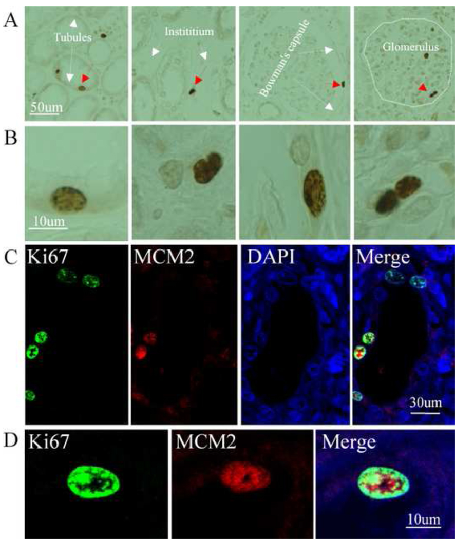
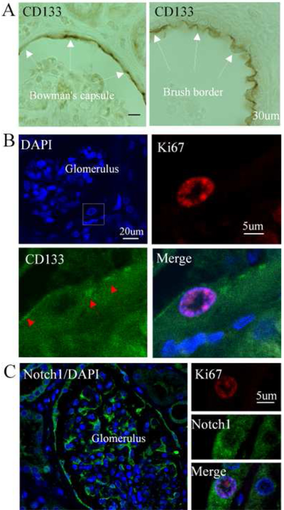
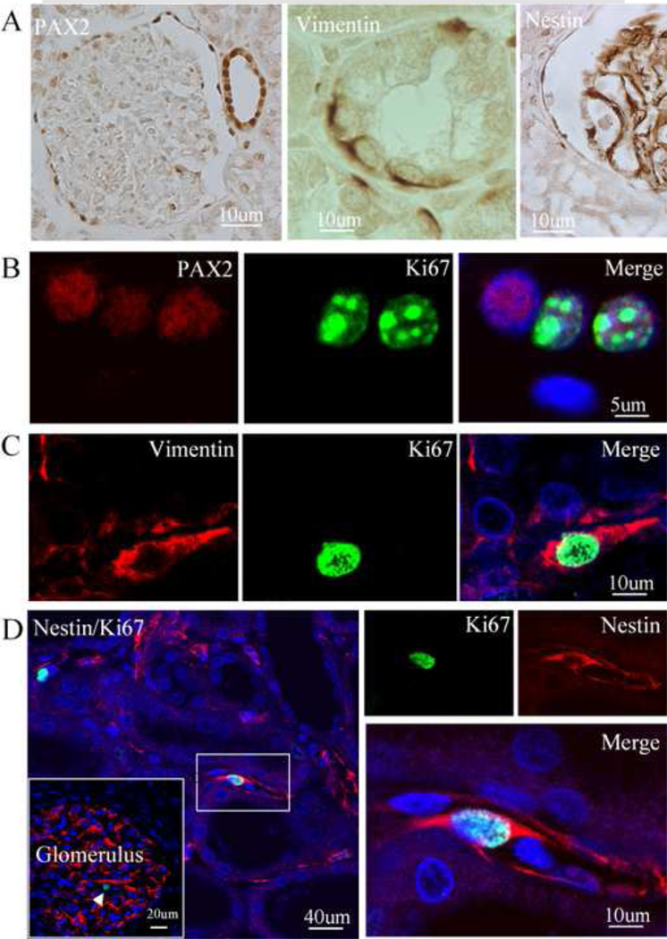
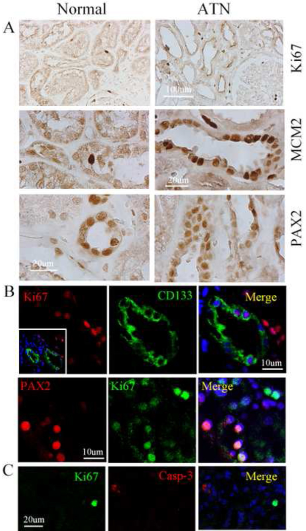
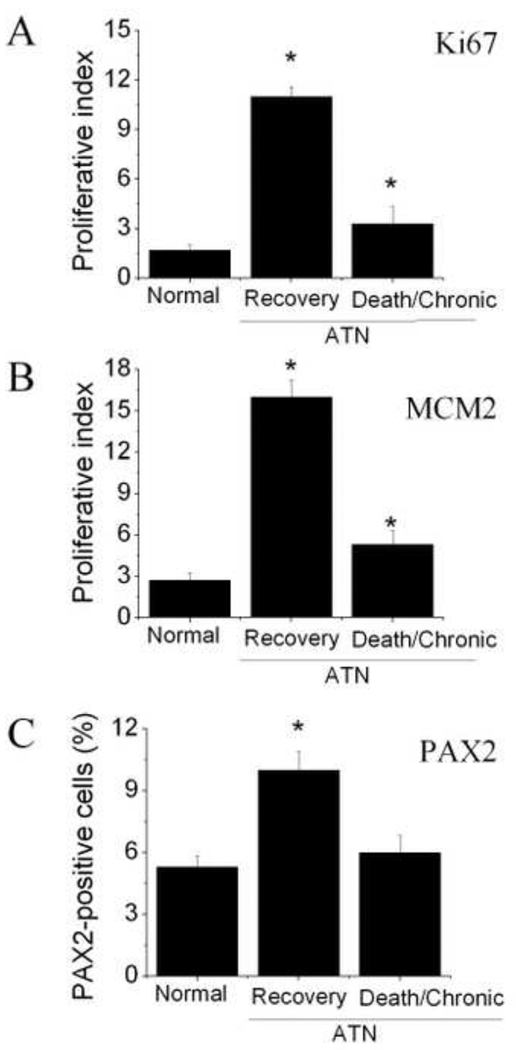
Animal studies indicate that adult renal stem/progenitor cells can undergo rapid proliferation in response to renal injury, but whether the same is true in humans is largely unknown. To examine the profile of renal stem/progenitor cells responsible for acute tubular necrosis in human kidney, double and triple immunostaining was performed using proliferative marker and stem/progenitor protein markers on sections from 10 kidneys with acute tubular necrosis and 4 normal adult kidneys. The immunopositive cells were recorded using 2-photon confocal laser scanning microscopy. We found that dividing cells were present in the tubules of the cortex and medulla, as well as the glomerulus in normal human kidney. Proliferative cells in the parietal layer of Bowman capsule expressed CD133, and dividing cells in the tubules expressed immature cell protein markers paired box gene 2, vimentin, and nestin. After acute tubular necrosis, Ki67-positive cells in the cortex tubules significantly increased compared with normal adult kidney. These Ki67-positive cells expressed CD133 and paired box gene 2, but not the cell death marker, activated caspase-3. In addition, the number of dividing cells increased significantly in patients with acute tubular necrosis who subsequently recovered, compared with patients with acute tubular necrosis who consequently developed protracted acute tubular necrosis or died. Our data suggest that renal stem/progenitor cells may reside not only in the parietal layer of Bowman capsule but also in the cortex and medulla in normal human kidney, and the proliferative capacity of renal stem/progenitor cells after acute tubular necrosis may be an important determinant of a patient's outcome.
Copyright © 2011 Elsevier Inc. All rights reserved.
Figures
References
-
- Poulsom R, Forbes SJ, Hodivala-Dilke K, Ryan E, Wyles S, Navaratnarasah S, Jeffery R, Hunt T, Alison M, Cook T, Pusey C, Wright NA. Bone marrow contributes to renal parenchymal turnover and regeneration. J Pathol. 2001;195:229–235. - PubMed
-
- Gupta S, Verfaillie C, Chmielewski D, Kim Y, Rosenberg ME. A role for extrarenal cells in the regeneration following acute renal failure. Kidney Int. 2002;62:1285–1290. - PubMed
-
- Anjos-Afonso F, Siapati EK, Bonnet D. In vivo contribution of murine mesenchymal stem cells into multiple cell-types under minimal damage conditions. J Cell Sci. 2004;117:5655–5664. - PubMed
-
- Szczypka MS, Westover AJ, Clouthier SG, Ferrara JL, Humes HD. Rare incorporation of bone marrow-derived cells into kidney after folic acid-induced injury. Stem Cells. 2005;23:44–54. - PubMed
Publication types
MeSH terms
Substances
Grants and funding
LinkOut - more resources
Full Text Sources
Medical
Research Materials
