Review
. 2011 Mar;34(3):134-42.
doi: 10.1016/j.tins.2011.01.002.

Development and cancer of the cerebellum

Affiliations
Review

Development and cancer of the cerebellum

Mary E Hatten et al. Trends Neurosci. 2011 Mar.

Abstract

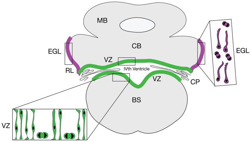
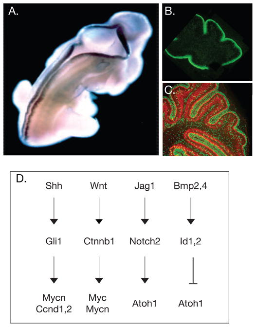
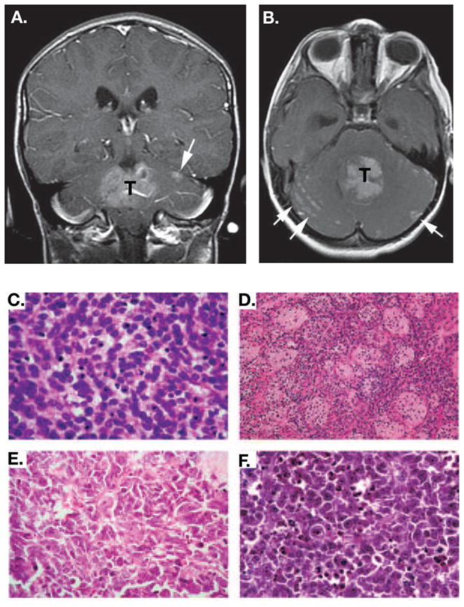
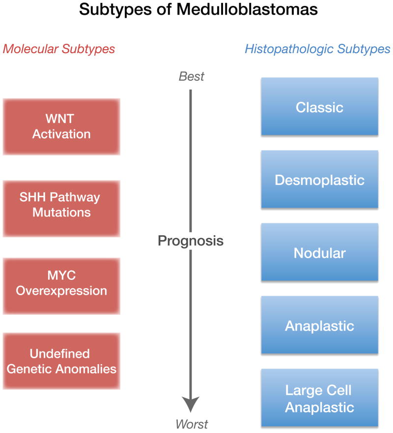
Medulloblastoma (MB) is the most common malignant pediatric brain tumor and is thought to arise from genetic anomalies in developmental pathways required for the normal maturation of the cerebellar cortex, notably developmental pathways for granule cell progenitor (GCP) neurogenesis. Over the past decade, a wide range of studies have identified genes and their regulators within signaling pathways, as well as noncoding RNAs, that have crucial roles in both normal cerebellar development and pathogenesis. These include the Notch, Wnt/β-catenin, bone morphogenic proteins (Bmp) and Sonic Hedgehog (Shh) pathways. In this review, we highlight the function of these pathways in the growth of the cerebellum and the formation of MB. A better understanding of the developmental origins of these tumors will have significant implications for enhancing the treatment of this important childhood cancer.


Conflict of interest statement

Disclosure statement

The authors have no conflict of interest related to this work.

Figures

Figure 1
Figure 1. Neurogenic Zones in Embryonic Cerebellar Histogenesis
In the embryonic cerebellar anlagen, the vast majority of cerebellar neurons, including Purkinje neurons (the major output neurons of the cerebellum), neurons of the cerebellar nuclei, more than half a dozen types of interneurons and cerebellar astroglia arise in the ventricular zone (VZ) lining the IVth ventricle (green). Different classes of cerebellar neurons are generated in a precise sequence, with neurons of the deep nuclei being generated first [18, 28], followed by Purkinje neurons and interneurons. Progenitors of the cerebellar nuclei migrate through the thickening wall of the anlage along the processes of radial glial cells to establish an external zone, which moves to a position deep to the Purkinje cells, as immature Purkinje neurons migrate away from the VZ and as GCPs migrate into the EGL, beginning at about E12 in the mouse [28]. GCPs emerge in a secondary neurogenic zone along the edge of the neuroepithelium in a zone called the rhombic lip (RL, purple). Proliferating precursors in this zone move onto the surface of the emerging anlage, where they form the EGL (purple), which gives rise to the GCPs, a subpopulation of neurons of the cerebellar nuclei [19, 21] and neurons of the lateral pontine nucleus in the brainstem [13]. Cells in the EGL (inset, purple cells) include mitotic figures and small cells with long migratory processes. Coronal view. BS, brainstem; CB, cerebellum; CP choroid plexus; EGL, external germinal layer; MB, midbrain; RL, rhombic lip.
Figure 2
Figure 2. Changing Patterns of Gene Expression and Signaling Pathways in Cerebellar Granule Cell Progenitor Expansion
(A) On E12.5 in the mouse, Atoh1/Math1 is expressed along the dorsal ridge of the spinal cord and in both the anterior and posterior rhombic lip. Adapted, with permission, from [76]. (B) In the postnatal cerebellar cortex, Atoh1/Math1 (green) is highly expressed in proliferating GCPS in the outer aspect of the EGL along the surface of the cerebellum (adapted, with permission, from [66])(C) As dividing GCPs (green) exit the cell cycle, they express p27Kip (red) (adapted, with permission, from [66]). (D) Four major pathways control GCP growth. The Shh pathway induces expression of the transcription factor Gli1, which up-regulates expression of cyclins (Ccnd D1, 2) and Mycn. Canonical Wnt signaling activates the b-catenin pathway, which up-regulates expression of both c-Myc and Mycn. Jag1 activates Notch2 signaling, which promotes proliferation and inhibits cell cycle exit, leading to a sustained expression of the bHLH transcription factor Atoh1/Math1. In contrast, Bmps are negative growth regulators. BMP2 and BMP4 induce expression of the transcription factor Id1,2, which inhibits Atoh1 expression, leading to cell cycle exit and neuronal differentiation.
Figure 3
Figure 3. Features of Human MB
MBs are diagnosed by imaging studies (MRI and CT scans) and histopathology. An axial (A) and a coronal (B) contrast-enhanced T1 weighted image are shown in which the enhancing tumor is labeled with T and metastases are evident in the subarachnoid space (white arrows). Adapted, with permission, from [86]. Histological variants of MB include: (C) Classic MB, which is typified by a field of small uniform cells with large nuclei, (D) The nodular/desmoplastic MB, which combines nodules of differentiated neurocytic cells with a low growth fraction and desmoplastic internodular zones of pleomorphic cells with a high growth fraction, (E) The anaplastic MB, which contains polymorphic cells with a high growth fraction; extensive apoptosis is also evident, and (F) Large cell MB, which contains groups of large cells with vesicular nuclei and a single nucleolus. Anaplasia is evident in other regions of this variant. Adapted, with permission,from [27].
Figure 4
Figure 4. Subtypes of MB are Defined by Histopathology or Molecular Features
Representation of two current classifications of MB subtypes, one based on histopathological features and the other on gene expression, and correlated outcome. The 5 subtypes of MBs defined by histopathology do not strictly overlap with the 4 subtypes defined by molecular analysis. At present, the most aggressive molecular subtype of MBs have undefined genetic anomalies.

References

    1. Bailey P, Cushing H. A Classification of the Tumors of the Glioma Group on a Histogenetic Basis With a Correlated Study of Prognosis. Lippincott; 1926.
    1. Palay SL, Chan-Palay S. The Cerebellar Cortex: cytology and organization. Springer-Verlag; 1974.
    1. Boyden ES, et al. Cerebellum-dependent learning: The Role of Multiple Plasticity Mechanisms. Annu Rev Neurosci. 2004;27:581–609. - PubMed
    1. De Zeeuw CI, Yeo CH. Time and tide in cerebellar memory formation. Current opinion in neurobiology. 2005;15:667–674. - PubMed
    1. du Lac S, et al. Learning and memory in the vestibulo-ocular reflex. Annu Rev Neurosci. 1995;18:409–441. - PubMed

Publication types