. 2011 Apr;59(4):536-48.
doi: 10.1016/j.yhbeh.2011.02.005. Epub 2011 Feb 18.

Chemosensory and hormone information are relayed directly between the medial amygdala, posterior bed nucleus of the stria terminalis, and medial preoptic area in male Syrian hamsters

Affiliations

Chemosensory and hormone information are relayed directly between the medial amygdala, posterior bed nucleus of the stria terminalis, and medial preoptic area in male Syrian hamsters

Laura E Been et al. Horm Behav. 2011 Apr.

Abstract

In many rodent species, including Syrian hamsters, the expression of appropriate social behavior depends critically on the perception and identification of conspecific odors. The behavioral response to these odors is mediated by a network of steroid-sensitive ventral forebrain nuclei including the medial amygdala (Me), posterior bed nucleus of the stria terminalis (BNST), and medial preoptic area (MPOA). Although it is well-known that Me, BNST, and MPOA are densely interconnected and each uniquely modulates odor-guided social behaviors, the degree to which conspecific odor information and steroid hormone cues are directly relayed between these nuclei is unknown. To answer this question, we injected the retrograde tracer, cholera toxin B (CTB), into the BNST or MPOA of male subjects and identified whether retrogradely-labeled cells in Me and BNST 1) expressed immediate early genes (IEGs) following exposure to male and/or female odors or 2) expressed androgen receptor (AR). Although few retrogradely-labeled cells co-localized with IEGs, a higher percentage of BNST- and MPOA-projecting cells in the posterior Me (MeP) expressed IEGs in response to female odors than to male odors. The percentage of retrogradely-labeled cells that expressed IEGs did not, however, differ between and female and male odor-exposed groups in the anterior Me (MeA), posterointermediate BNST (BNSTpi), or posteromedial BNST (BNSTpm). Many retrogradely-labeled cells co-localized with AR, and a higher percentage of retrogradely-labeled MeP and BNSTpm cells expressed AR than retrogradely-labeled MeA and BNSTpi cells, respectively. Together, these data demonstrate that Me, BNST, and MPOA interact as a functional circuit to process sex-specific odor cues and hormone information in male Syrian hamsters.


Figures

Figure 1
Figure 1
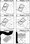
Counting domains for analysis of cholera toxin B (CTB), immediate early genes (Fos, EGR-1) and androgen receptor (AR). The total number of single- and double-labeled cells within the specified counting domains (grey rectangles) were counted and summed across two sections for the anterior medial amygdala (MeA; A and B), posterior medial amygdala (MeP; C and D), and posterointermediate and posteromedial bed nucleus of the stria terminalis (BNSTpi and BNSTpm; E and F). Illustration modified from hamster brain atlas (Morin and Wood, 2001). ACo, anterior cortical amygdala; BLA, basolateral amygdala; BLP, posterior basolateral amygdala; BMA, basomedial amygdala; CeA, central amygdala; f, fornix; GP, globus pallidus; I, intercalated amygdala; ic, internal capsule; LPO, lateral preoptic area; ot, optic tract; MPN, medial preoptic nucleus; MPO, medial preoptic area; PLCo posterolateral cortical amygdala; sm, stria medullaris; St, stria terminalis.
Figure 2
Figure 2
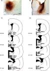
Verification of cholera toxin B (CTB) injections. Photomicrographs depict typical deposition of CTB in A) the medial preoptic area (MPOA) and B) the posterior bed nucleus of the stria terminalis (BNST). Reconstruction of largest (light grey) and smallest (dark grey) CTB injections (numbers represent distance posterior to bregma) through the rostral-caudal extent of (C) MPOA and (D) BNST. Scale bars = 100 μm.
Figure 3
Figure 3
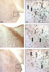
Co-localization of cholera toxin B (CTB) and immediate early genes (Fos, EGR-1). Photomicrographs from representative sections immunoreactive for CTB and Fos in A) the medial amygdala (Me) and B) the posterior bed nucleus of the stria terminalis (BNST), and C) from a representative section immunoreactive for CTB and EGR-1 in Me. Images to right provide higher magnifications of areas included in the boxes. Cells with brown cytoplasmic staining (white arrows) are CTB+, cells with black nuclear staining (black arrows) are Fos+ (A and B) or EGR-1+ (C), and cells with black nuclear staining surrounded by brown cytoplasmic staining (grey arrows) are Fos/CTB+ (A and B) or EGR-1/CTB+ (C). Scale bars = 100 μm and 25 μm for lower and higher magnifications, respectively.
Figure 4
Figure 4
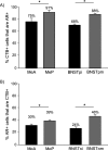
Percentages of Fos/CTB double-labeled cells in males with medial preoptic area (MPOA) injections. A) Total percentage of CTB+ cells that were also Fos+ in the anterior medial amygdala (MeA), posterior medial amygdala (MeP), posterointermediate bed nucleus of the stria terminalis (BNSTpi), and posteromedial bed nucleus of the stria terminalis (BNSTpm). B) Total percentage of Fos+ that were also CTB+ in MeA, MeP, BNSTpi, and BNSTpm. Error bars represent standard errors of mean group percentages, * reflects significant differences between brain areas, P < 0.05 (z-test for independent proportions, αfw = 0.05).
Figure 5
Figure 5
Percentages of Fos/CTB double-labeled cells in males with posterior bed nucleus of the stria terminalis (BNST) injections. A) Total percentage of CTB+ cells that were also Fos+ in the anterior medial amygdala (MeA) and posterior medial amygdala (MeP). B) Total percentage of Fos+ that were also CTB+ in MeA and MeP. Error bars represent standard errors of mean group percentages, * reflects significant differences between brain areas, P < 0.05 (z-test for independent proportions, αfw = 0.05).
Figure 6
Figure 6
Co-localization of cholera toxin B (CTB) and androgen receptor (AR). Photomicrographs from representative sections immunoreactive for CTB and AR in A) the medial amygdala (Me) and B) the posterior bed nucleus of the stria terminalis (BNST). Images to right provide higher magnifications of areas included in the boxes. Cells with brown cytoplasmic staining (white arrows) are CTB+, cells with black nuclear staining (black arrows) are AR+, and cells with black nuclear staining surrounded by brown cytoplasmic staining (grey arrows) are AR/CTB+. Scale bars = 100 μm and 25 μm for lower and higher magnifications, respectively.
Figure 7
Figure 7
Percentages of AR/CTB double-labeled cells in males with medial preoptic area (MPOA) injections. A) Total percentage of CTB+ cells that were also AR+ in the anterior medial amygdala (MeA), posterior medial amygdala (MeP), posterointermediate bed nucleus of the stria terminalis (BNSTpi) and posteromedial bed nucleus of the stria terminalis (BNSTpm). B) Total percentage of AR+ cells that were also CTB+ in MeA, MeP, BNSTpi, and BNSTpm. Error bars represent standard errors of mean group percentages, * reflects significant differences between brain areas, P < 0.05 (z-test for independent proportions, αfw = 0.05).
Figure 8
Figure 8
Percentages of AR/CTB double-labeled cells in males with posterior bed nucleus of the stria terminalis (BNST) injections. A) Total percentage of CTB+ cells that were also AR+ in the anterior medial amygdala (MeA) and posterior medial amygdala (MeP). B) Total percentage of AR+ cells that were also CTB+ in MeA and MeP. Error bars represent standard errors of mean group percentages, * reflects significant differences between brain areas, P < .05 (z-test for independent proportions, αfw = 0.05).
Figure 9
Figure 9
Summary of chemosensory and steroid-sensitive projections to medial preoptic area (MPOA) and posterior bed nucleus of the stria terminalis (BNST). A) A low percentage of cells that respond to female odors (filled circles) or male odors (open circles) project to MPOA (left) or BNST (right). In the posterior medial amygdala (MeP), however, a higher percentage of female-odor responsive cells project to BNST and/or MPOA than do male odor-responsive cells. B) A high percentage of androgen receptor (AR)-bearing cells (squares) project to MPOA (left) and BNST (right). Specifically, more AR+ cells in MeP than in the anterior medial amygdala (MeA) project to MPOA and/or BNST; similarly, more AR+ cells in the posteromedial BNST (BNSTpm) than in the posterointermediate BNST (BNSTpi) project to MPOA. Solid arrows indicate afferent projections; dashed arrows represent direct olfactory projections.

References

    1. Ballard CL, Wood RI. Partner preference in male hamsters: steroids, sexual experience and chemosensory cues. Physiol Behav. 2007;91:1–8. - PMC - PubMed
    1. Baum MJ, Everitt BJ. Increased expression of c-fos in the medial preoptic area after mating in male rats: role of afferent inputs from the medial amygdala and midbrain central tegmental field. Neuroscience. 1992;50:627–46. - PubMed
    1. Baum MJ, Kelliher KR. Complementary roles of the main and accessory olfactory systems in mammalian mate recognition. Annu Rev Physiol. 2009;71:141–60. - PubMed
    1. Been L, Petrulis A. Society for Neuroscience. Washington, D.C.: 2008. The role of the posterior bed nucleus of the stria terminalis in opposite-sex odor preference and sexual odor processing in male Syrian hamsters. - PubMed
    1. Been L, Petrulis A. Lesions of the posterior bed nucleus of the stria terminalis eliminate opposite-sex odor preference and delay copulation in male Syrian hamsters: role of odor volatility and sexual experence. Eur J Neurosci. 2010a - PMC - PubMed

Publication types

Substances