. 2011 Mar 1;25(5):485-98.
doi: 10.1101/gad.2019811. Epub 2011 Feb 11.

EZH1 and EZH2 cogovern histone H3K27 trimethylation and are essential for hair follicle homeostasis and wound repair

Affiliations

EZH1 and EZH2 cogovern histone H3K27 trimethylation and are essential for hair follicle homeostasis and wound repair

Elena Ezhkova et al. Genes Dev. .

Abstract

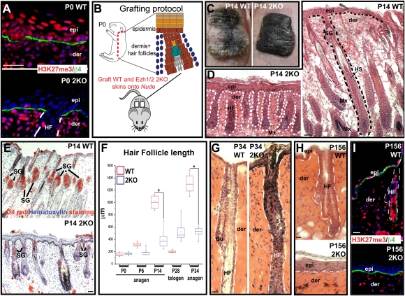
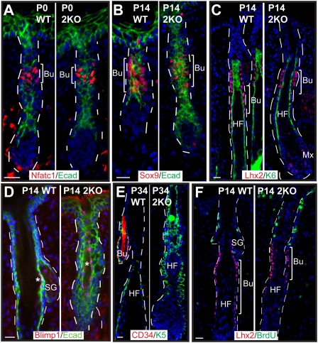
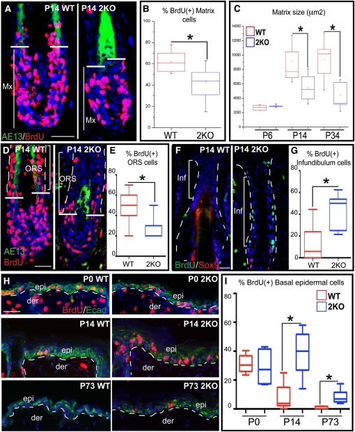
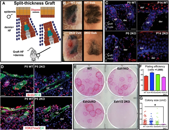
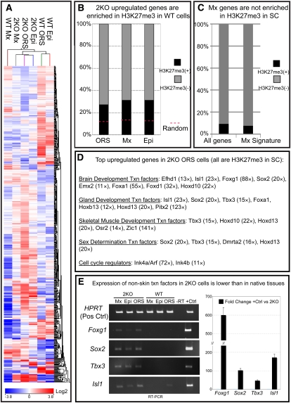
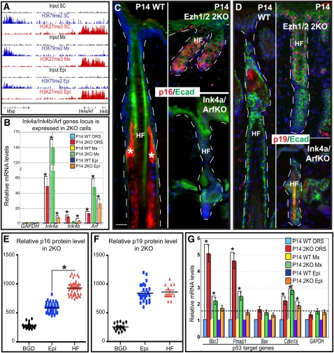
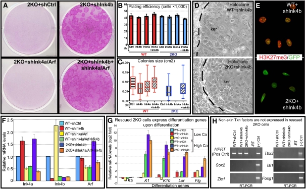
Polycomb protein group (PcG)-dependent trimethylation on H3K27 (H3K27me3) regulates identity of embryonic stem cells (ESCs). How H3K27me3 governs adult SCs and tissue development is unclear. Here, we conditionally target H3K27 methyltransferases Ezh2 and Ezh1 to address their roles in mouse skin homeostasis. Postnatal phenotypes appear only in doubly targeted skin, where H3K27me3 is abolished, revealing functional redundancy in EZH1/2 proteins. Surprisingly, while Ezh1/2-null hair follicles (HFs) arrest morphogenesis and degenerate due to defective proliferation and increased apoptosis, epidermis hyperproliferates and survives engraftment. mRNA microarray studies reveal that, despite these striking phenotypic differences, similar genes are up-regulated in HF and epidermal Ezh1/2-null progenitors. Featured prominently are (1) PcG-controlled nonskin lineage genes, whose expression is still significantly lower than in native tissues, and (2) the PcG-regulated Ink4a/Inkb/Arf locus. Interestingly, when EZH1/2 are absent, even though Ink4a/Arf/Ink4b genes are fully activated in HF cells, they are only partially so in epidermal progenitors. Importantly, transduction of Ink4b/Ink4a/Arf shRNAs restores proliferation/survival of Ezh1/2-null HF progenitors in vitro, pointing toward the relevance of this locus to the observed HF phenotypes. Our findings reveal new insights into Polycomb-dependent tissue control, and provide a new twist to how different progenitors within one tissue respond to loss of H3K27me3.


Figures

Figure 1.
Figure 1.
Arrested morphogenesis and progressive degeneration of HFs and SGs lacking EZH1 and EZH2. (A) Immunofluorescence confirms the absence of H3K27me3 in P0 Ezh1/2 double-knockout (2KO) skin epithelium. Tissue sections are counterstained for β4 integrin (green) and chromatin (DAPI, blue). (Epi) Epidermis; (der) dermis. (B) Schematic of full-thickness grafting protocol (Kaufman et al. 2003) used to examine postnatal consequences of Ezh1/2 ablation. (C) Absence of surface hair in double-knockout versus wild-type (WT) skin grafts. (D) Histological analysis reveals that HFs of double-knockout grafts are short, and lack hair shafts (HS). (Mx) Matrix. (E) Oil-red-O stains lipids and confirms impaired SG formation in Ezh1/2 double-knockout skins. (F) Quantification of HF lengths at times and hair cycle stages indicated. Note that double-knockout HFs arrest in the first growth phase of the hair cycle. (G) The old hair and surrounding bulge (Bu) SC compartment are evident in wild-type but not double-knockout HFs by P34. (H) P156 grafts reveal that wild-type HFs have undergone two hair cycles, but double-knockout skin has lost all HFs. Note hyperthickening of epidermis in double-knockout skin. (I) Immunofluorescence confirming that P156 double-knockout epidermis lacks H3K27me3. Bars: A,G–I, 30 μm; D,E, 90 μm.
Figure 2.
Figure 2.
Cells expressing HF-SC and SG-SC markers are still present in Ezh1/2 double-knockout skin. (A–E) Immunofluorescence microscopy of sections from P0 (A), P14 (B–D), or P34 (E) skin grafts analyzed for temporal and spatial expression of SC markers. Color-coding is according to the secondary antibodies used. Blimp1 is a marker of SG-SCs; Nfatc1, Lhx2, Sox9, and CD34 are HF-SC markers. (F) A short (4-h) BrdU pulse followed by immunofluorescence analysis shows that, similar to wild type, none of Lhx2+ SCs are BrdU-labeled. In contrast, the cells above the bulge are hyperproliferative in double-knockout compared with wild-type skin. Bar, 30 μm.
Figure 3.
Figure 3.
Loss of EZH1/2 results in reduced proliferation of HF ORS and matrix, but hyperproliferation in infundibulum and epidermis. (A–E) A short (4-h) BrdU pulse was administered to engrafted mice prior to processing the engrafted tissue for anti-BrdU and hair keratin (AE13) immunofluorescence microscopy and quantitative analyses. Note reduction in proliferation in the matrix (Mx), hair shaft precursors (cells above horizontal bars and internal to or including AE13+ layer) and ORS (cells above horizontal bars and external to and excluding AE13+ layer) in double-knockout (2KO) versus wild-type (WT) skins. Note also that overall matrix size of double-knockout HFs was normal at P6, but markedly diminished by P14 and P34 post-engraftment. (F–I) Similar analyses as in A–E, except on double-knockout infundibulum and interfollicular epidermis. Note hyprerproliferation in both compartments by P14. By P73, double-knockout grafts still show more proliferation than wild type, but overall proliferative levels in the epidermis decline with age. Bar, 30 μm.
Figure 4.
Figure 4.
Double-knockout HFs are defective in proliferation even upon wound-induced stimulation. (A) Schematic of split-thickness grafting protocol. The ability of HF cells to migrate out and re-epithelialize the epidermis is largely dependent on functional bulge SCs, although wounds will eventually close by Nude mouse epidermal migration (Nowak et al. 2008). Male dermis + HFs are engrafted onto female Nude mice to distinguish the two events by Y-chromosome in situ hybridization. (B) Six weeks after engraftment, double-knockout split thickness grafts finally close their wounds, a feat accomplished by their wild-type counterparts in 2 wk. (C) At P0, epidermis is completely missing in split-thickness grafts. By P14, re-epithelialization of wild-type grafts is accomplished largely by HF migration (Y-chr+). In double-knockout grafts, re-epithelialization, still in progress, is exclusively by Nude host-derived epidermal migration. (D) A short BrdU pulse administered 5 d post-engraftment shows lack of a proliferative response by the H3K27me3-deficient HFs of double-knockout skin. (E–G) Double-knockout HF progenitors are unable to proliferate in vitro. (E) Rhodamine B staining of primary mouse HF cultures after 3 wk in enriched media. Note that only doubly targeted cells fail to form visible colonies. Quantifications show that plating efficiencies are comparable but colonies simply fail to grow in the absence of H3K27me3. Bar, 30 μm.
Figure 5.
Figure 5.
Ablation of Ezh1/2 leads to the up-regulation of a common subset of genes in three different skin progenitors irrespective of their lineage and response to deregulation of PcG-mediated repression. (A) Heat map and clustering analyses reveal that many genes differentially expressed in double-knockout progenitors of ORS, matrix (Mx), and epidermis (Epi) cocluster together. Notably, genes up-regulated in double-knockout ORS progenitors do not cocluster with wild-type matrix or wild-type epidermis cells, indicating that double-knockout progenitors are not induced to progress toward HF (matrix) or epidermal lineages. (B) The Ezh1/2 up-regulated genes of each skin progenitor type was compared against ChIP-seq data mapping H3K27me3 (PcG-repressive) and H3K79me2 (active transcription) marks in the chromatin of postnatal bulge SCs, matrix TA cells, or epidermal basal progenitors. Whole-scale comparative analysis reveals that one-third of genes up-regulated in double-knockout ORS, matrix, or epidermis are targeted by H3K27me3 repression in wild-type progenitors. (C) Genes that are normally activated in wild-type matrix cells are not preferentially repressed by H3K27me3 histone mark in SCs, explaining why genes controlling matrix lineage determination are not preferentially up-regulated when H3K27me3 marks are removed from ORS stem cell progenitors. (D) Top up-regulated genes in double-knockout ORS cells that are regulated by H3K27me3 in wild-type ORS bulge SCs. Fold up-regulation is given in parentheses. (E) Real-time PCR reveals that ectopic activation of genes encoding nonskin lineage transcription factors in all three skin progenitors is significantly lower compared with their levels within native tissues (+Ctrl). Quantifications are shown at right.
Figure 6.
Figure 6.
The Ink4b/Ink4a/Arf gene locus is targeted by EZH1/2-dependent H3K27me3 in skin progenitors, but activation levels upon Ezh1/2 ablation differ markedly among skin progenitor populations. (A) ChIP-seq data reveal that the Ink4b/Ink4a/Arf gene locus is targeted by H3K27me3 in bulge SCs (SC)/ORS, matrix (Mx), and basal epidermal (Epi) cells. ChIP-seq for the H3K79me2 histone mark confirms that these H3K27me3-decorated genes are in a repressed state. The housekeeping gene Mtap is shown here as a control. (B) RT-qPCR reveals that Ink4b, Ink4a, and Arf genes are all up-regulated in double-knockout skin progenitors relative to their wild-type counterparts, but the levels of induction vary dramatically among progenitor populations. (C,D) Immunofluorescence confirms variation in the induction of p16 (Ink4a) and p19 (Arf) by epidermal versus HF progenitors. Ink4a/Arf knockout tissue was used to control for antibody specificity. Note elevated nuclear (active) p16 and p19 labeling in double-knockout progenitors, not seen in wild type. Wild type only shows cytoplasmic staining (asterisk) of terminally differentiated HF lineages. (E,F) Quantifications of relative protein levels shows that p16 is significantly lower in double-knockout epidermis versus HF, while p19 levels are comparable. (G) P19 stabilizes p53, whose transcriptional activity can be monitored by activation of its target genes: Bbc3(PUMA; proapoptotic protein), Pmaip1 (NOXA; proapoptotic protein), and Bax and Cdkn1a (p21). Note that, despite comparable expression of p19 protein in double-knockout epidermis and HFs, p53 target proapoptotic genes are significantly more activated in the HF than epidermal progenitors. Bar, 30 μm.
Figure 7.
Figure 7.
shRNA-mediated down-regulation of Ink4b/Ink4a/Arf expression restores proliferation of Ezh1/2 double-knockout HF progenitors in vitro. (A–D) P0 wild-type and double-knockout HF cells were cultured and transduced with lentiviral expression vectors harboring shRNA hairpins that target Ink4a/Arf (shared region) and/or Ink4b. Rhodamine B staining (A) and quantification (B,C) revealed that double-knockout cells expressing control (scrambled) shRNA or shRNA targeting Ink4a/Arf fail to form visible colonies 4 wk after plating, in contrast to double-knockout cells expressing Ink4b and (optimally) Ink4b + Ink4a/Arf. Plating efficiencies (B) were comparable, but proliferation (C) and colony morphologies (D) were significantly enhanced when the locus was repressed. (E) Immunofluorescence confirms that the shInk4b transduced (GFP+) double-knockout cells still lack the H3K27me3 mark in contrast to transduced wild-type cells. (F) Efficiencies of RNAi knockdown of Ink4b and Ink4a/Arf genes as analyzed by RT-qPCR on FACS-isolated populations 4 wk after transduction. (G) Double-knockout + shInk4b cells are able to induce terminal differentiation genes keratin 1 (K1), keratin 10 (K10), loricrin (Lor), and filaggrin (Flg) upon a shift from low to high calcium. Analysis is by RT-qPCR. (H) Genes encoding transcription factors of nonskin lineages are not expressed in cultured HF progenitors that can grow following rescue of the Ink4a/Arf/Ink4b locus.

References

    1. Barrandon Y, Green H 1987. Three clonal types of keratinocyte with different capacities for multiplication. Proc Natl Acad Sci 84: 2302–2306 - PMC - PubMed
    1. Barski A, Cuddapah S, Cui K, Roh TY, Schones DE, Wang Z, Wei G, Chepelev I, Zhao K 2007. High-resolution profiling of histone methylations in the human genome. Cell 129: 823–837 - PubMed
    1. Blanpain C, Fuchs E 2009. Epidermal homeostasis: a balancing act of stem cells in the skin. Nat Rev Mol Cell Biol 10: 207–217 - PMC - PubMed
    1. Blanpain C, Lowry WE, Geoghegan A, Polak L, Fuchs E 2004. Self-renewal, multipotency, and the existence of two cell populations within an epithelial stem cell niche. Cell 118: 635–648 - PubMed
    1. Boyer LA, Plath K, Zeitlinger J, Brambrink T, Medeiros LA, Lee TI, Levine SS, Wernig M, Tajonar A, Ray MK, et al. 2006. Polycomb complexes repress developmental regulators in murine embryonic stem cells. Nature 441: 349–353 - PubMed

Publication types

MeSH terms

Substances