. 2011 Apr;77(7):2445-57.
doi: 10.1128/AEM.02643-10. Epub 2011 Feb 11.

Contribution of rapid evolution of the luxR-luxI intergenic region to the diverse bioluminescence outputs of Vibrio fischeri strains isolated from different environments

Affiliations

Contribution of rapid evolution of the luxR-luxI intergenic region to the diverse bioluminescence outputs of Vibrio fischeri strains isolated from different environments

Jeffrey L Bose et al. Appl Environ Microbiol. 2011 Apr.

Abstract

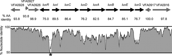
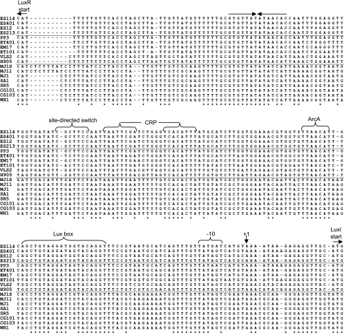
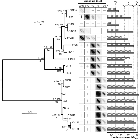
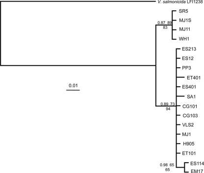
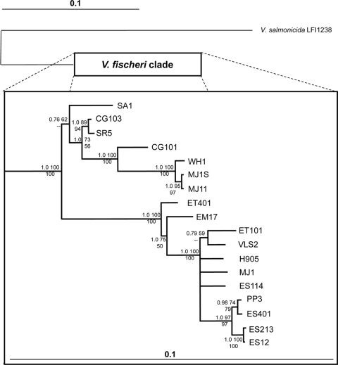
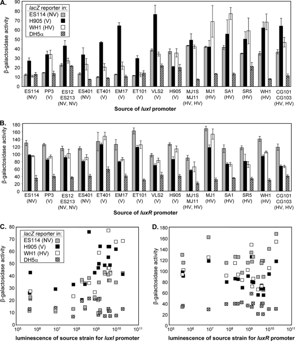
Vibrio fischeri serves as a valuable model of bacterial bioluminescence, its regulation, and its functional significance. Light output varies more than 10,000-fold in wild-type isolates from different environments, yet dim and bright strains have similar organization of the light-producing lux genes, with the activator-encoding luxR divergently transcribed from luxICDABEG. By comparing the genomes of bright strain MJ11 and the dimmer ES114, we found that the lux region has diverged more than most shared orthologs, including those flanking lux. Divergence was particularly high in the intergenic sequence between luxR and luxI. Analysis of the intergenic lux region from 18 V. fischeri strains revealed that, with one exception, sequence divergence essentially mirrored strain phylogeny but with relatively high substitution rates. The bases conserved among intergenic luxR-luxI sequences included binding sites for known regulators, such as LuxR and ArcA, and bases of unknown significance, including a striking palindromic repeat. By using this collection of diverse luxR-luxI regions, we found that expression of P(luxI)-lacZ but not P(luxR)-lacZ transcriptional reporters correlated with the luminescence output of the strains from which the promoters originated. We also found that exchange of a small stretch of the luxI-luxR intergenic region between two strains largely reversed their relative brightness. Our results show that the luxR-luxI intergenic region contributes significantly to the variable luminescence output among V. fischeri strains isolated from different environments, although other elements of strain backgrounds also contribute. Moreover, the lux system appears to have evolved relatively rapidly, suggesting unknown environment-specific selective pressures.


Figures

FIG. 1.
FIG. 1.
Conservation of lux and flanking regions between V. fischeri strains ES114 and MJ11. The percent identity is shown for aligned nucleotides (on the y axis of the inset graph) and for encoded proteins as the percent amino acid (AA) identity below the corresponding gene. The black and gray arrows indicate the orientation of lux genes or flanking genes, respectively. Graphic representation of the percent nucleotide identity was generated with VISTA (40) and the LAGAN alignment program (9) using default settings. An asterisk marks an unshaded region between luxI and luxR where a window in the alignment falls below the 70% identity cutoff. Conservation of regions flanking the lux genes was typical for that extending several kilobases in each direction (data not shown).
FIG. 2.
FIG. 2.
Alignment of lux intergenic regions from diverse V. fischeri strains. Horizontal dashed lines separate strains with luminescence on plates classified as nonvisible (ES114, ES401, ES12, and ES213), visible (PP3, ET401, EM17, ET101, VLS2, and H905), or highly visible (MJ1S, MJ11, MJ1, SA1, SR5, CG101, CG103, and WH1). Asterisks mark bases conserved across all strains. Labeled arrows mark the translational start sites for LuxI and LuxR (on the reverse strand). The transcriptional start site for luxICDABEG determined in ES114 (16) is marked +1 and with a vertical arrow, and the corresponding −10 promoter element is indicated. Core consensus sequences for CRP, ArcA, and LuxR binding are shown and are discussed further in the text. A pair of arrows above the top row of the sequences indicates the position of an inverted repeat. The region labeled the site-directed switch was engineered such that this sequence in ES114 was placed in MJ1S to generate strain JB7 and, likewise, switched from MJ1S into ES114 to generate strain JB3.
FIG. 3.
FIG. 3.
Relatedness of the luxR-luxI intergenic sequence in V. fischeri isolates. The topology represents the 50% majority rule consensus tree generated from a Bayesian analysis. Statistical support of each node is given at the particular node based on three values: Bayesian posterior probability (upper left); ML bootstrap support values (1,000 pseudoreplicates) (upper right); MP bootstrap support values (1,000 pseudoreplicates) (lower center). Dashes indicate clade support of <50% for 1,000 MP bootstrap pseudoreplicates. The tree was mid-point rooted. Bar, 0.1 substitutions per site. Patches were grown overnight on SWTO at 24°C, and the negative images for given exposures are shown. A + or − indicates that luminescence was too high or low to see clearly, respectively. Horizontal bars display the luminescence per OD595 unit of each strain grown on plates for 18 h and resuspended (hatched bars) or grown in broth with peak luminescence reported (solid bars). Dashed vertical lines indicate approximate cutoffs that were used to distinguish strains that produced nonvisible, visible, or highly visible luminescence on SWTO plates.
FIG. 4.
FIG. 4.
Bayesian phylogeny of the intergenic glpA-fdhA region among V. fischeri isolates. The topology was generated as described for Fig. 3, and the statistical support values given at each node were calculated and are placed in order as for Fig. 3. The tree was rooted with the corresponding sequence from V. salmonicida LFI1238. Bar, 0.01 substitutions per site. This reconstruction's low resolution is a result of low sequence diversity among isolates.
FIG. 5.
FIG. 5.
Bayesian phylogeny of concatenated recA-katA-mdh sequences among V. fischeri isolates. The topology was generated as described for Fig. 3, and the statistical support values given at each node were calculated and placed in order as for Fig. 3. The tree was rooted with the corresponding sequences from V. salmonicida LFI1238. Bars, 0.1 substitutions per site; the V. fischeri clade was expanded in order to more easily view fine-scale topological resolution among all isolates.
FIG. 6.
FIG. 6.
Activity of lux promoter reporters. The β-galactosidase activities (in Miller units) from specific PluxI-lacZ and PluxR-lacZ constructs are shown for reporters carried in V. fischeri strains ES114 (gray), H905 (black), and WHI (open) as well as in E. coli DH5α (hatched lines). The luminescence class of the V. fischeri strains (Table 1) is indicated in parentheses as NV, V, or HV. (A and B) The strain(s) corresponding to the promoter sequence in PluxI-lacZ and PluxR-lacZ constructs, respectively, and these are arranged (left to right) in the same order as the strains shown in Fig. 3 (top to bottom). (C and D) Reporter activity is plotted as a function of the luminescence of the source strain(s) for PluxI-lacZ and PluxR-lacZ promoters, respectively. Data from different experiments with treatments in common and with similar results were pooled, and averages are reported (panels A and B also show standard errors [n = 3]).
FIG. 7.
FIG. 7.
Exchanging a short stretch of the luxI-luxR intergenic region between ES114 and MJ1S largely reverses their relative brightness. Luminescence per OD595 unit is shown for cultures of ES114, JB3, MJ1S, and JB7 grown in SWTO broth at 24°C with shaking (200 rpm). Data are the average peak luminescence levels per OD595 unit, with standard errors (n = 2).

Similar articles

Cited by

References

    1. Adin, D. M., K. L. Visick, and E. V. Stabb. 2008. Identification of a cellobiose utilization gene cluster with cryptic beta-galactosidase activity in Vibrio fischeri. Appl. Environ. Microbiol. 74:4059-4069. - PMC - PubMed
    1. Altschul, S. F., W. Gish, W. Miller, E. W. Myers, and D. J. Lipman. 1990. Basic local alignment search tool. J. Mol. Biol. 215:403-410. - PubMed
    1. Antunes, L. C., R. B. Ferreira, C. P. Lostroh, and E. P. Greenberg. 2008. A mutational analysis defines Vibrio fischeri LuxR binding sites. J. Bacteriol. 190:4392-4397. - PMC - PubMed
    1. Ast, J. C., H. Urbanczyk, and P. V. Dunlap. 2009. Multi-gene analysis reveals previously unrecognized phylogenetic diversity in Aliivibrio. Syst. Appl. Microbiol. 32:379-386. - PubMed
    1. Boettcher, K. J., and E. G. Ruby. 1990. Depressed light emission by symbiotic Vibrio fischeri of the sepiolid squid Euprymna scolopes. J. Bacteriol. 172:3701-3706. - PMC - PubMed

Publication types

Associated data

LinkOut - more resources