

Amount but not pattern of protective sensory stimulation alters recovery after permanent middle cerebral artery occlusion
- PMID: 21317269
- PMCID: PMC3141731
- DOI: 10.1161/STROKEAHA.110.607135
Amount but not pattern of protective sensory stimulation alters recovery after permanent middle cerebral artery occlusion
Abstract
Background and purpose: Using a rodent model of ischemia (permanent middle cerebral artery occlusion), our laboratory previously demonstrated that 4.27 minutes of patterned single-whisker stimulation delivered over 120 minutes can fully protect from impending damage when initiated within 2 hours of permanent middle cerebral artery occlusion ("early"). When initiated 3 hours postpermanent middle cerebral artery occlusion ("late"), stimulation resulted in irreversible damage. Here we investigate the effect of altering pattern, distribution, or amount of stimulation in this model.
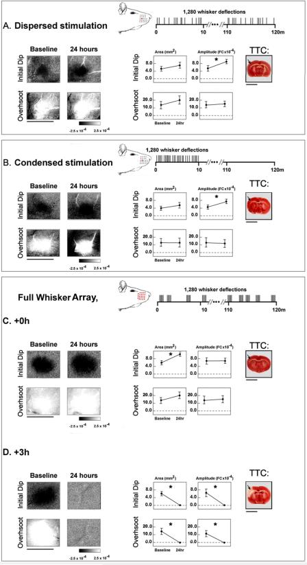
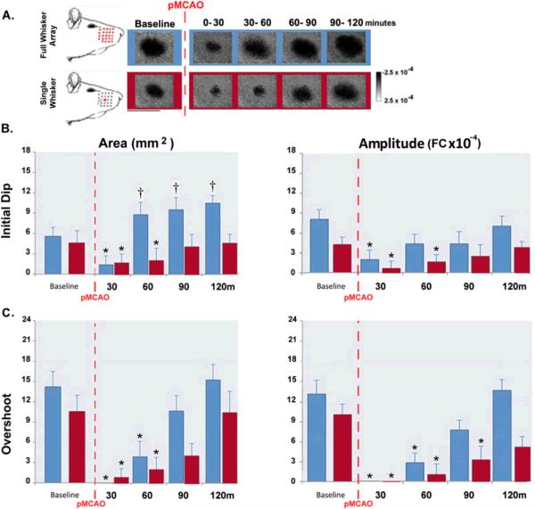
Methods: We assessed the cortex using functional imaging and histological analysis with altered stimulation treatment protocols. In 2 groups of animals we administered the same number of whisker deflections but in a random rather than patterned fashion distributed either over 120 minutes or condensed into 10 minutes postpermanent middle cerebral artery occlusion. We also tested increased (full-whisker array versus single-whisker) stimulation.
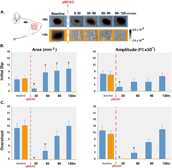
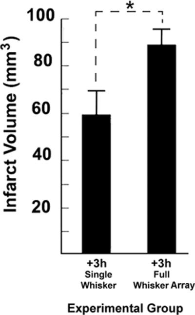
Results: Early random whisker stimulation (condensed or dispersed) resulted in protection equivalent to early patterned stimulation. Early full-whisker array patterned stimulation also resulted in complete protection but promoted faster recovery. Late full-whisker array patterned stimulation, however, resulted in loss of evoked function and infarct volumes larger than those sustained by single-whisker counterparts.
Conclusions: When induced early on after ischemic insult, stimulus-evoked cortical activity, irrespective of the parameters of peripheral stimulation that induced it, seems to be the important variable for neuroprotection.
Figures
References
-
- Lloyd-Jones D, Adams RJ, Brown TM, Carnethon M, Dai S, De Simone G, Ferguson TB, Ford E, Furie K, Gillespie C, Go A, Greenlund K, Haase N, Hailpern S, Ho PM, Howard V, Kissela B, Kittner S, Lackland D, Lisabeth L, Marelli A, McDermott MM, Meigs J, Mozaffarian D, Mussolino M, Nichol G, Roger VL, Rosamond W, Sacco R, Sorlie P, Roger VL, Thom T, Wasserthiel-Smoller S, Wong ND, Wylie-Rosett J. Heart disease and stroke statistics--2010 update: A report from the american heart association. Circulation. 2010;121:e46–e215. - PubMed
-
- Tamura A, Graham DI, McCulloch J, Teasdale GM. Focal cerebral ischaemia in the rat: 1. Description of technique and early neuropathological consequences following middle cerebral artery occlusion. J Cereb Blood Flow Metab. 1981;1:53–60. - PubMed
-
- Brint S, Jacewicz M, Kiessling M, Tanabe J, Pulsinelli W. Focal brain ischemia in the rat: Methods for reproducible neocortical infarction using tandem occlusion of the distal middle cerebral and ipsilateral common carotid arteries. J Cereb Blood Flow Metab. 1988;8:474–485. - PubMed
-
- Wang-Fischer Y. Manual of stroke models in rats. CRC Press; Boca Raton: 2009.
Publication types
MeSH terms
Grants and funding
LinkOut - more resources
Full Text Sources
