. 2011 Apr 8;286(14):12371-80.
doi: 10.1074/jbc.M110.182931. Epub 2011 Feb 11.

Mapping of the SecA·SecY and SecA·SecG interfaces by site-directed in vivo photocross-linking

Affiliations

Mapping of the SecA·SecY and SecA·SecG interfaces by site-directed in vivo photocross-linking

Sanchaita Das et al. J Biol Chem. .

Abstract

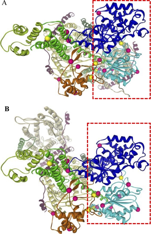
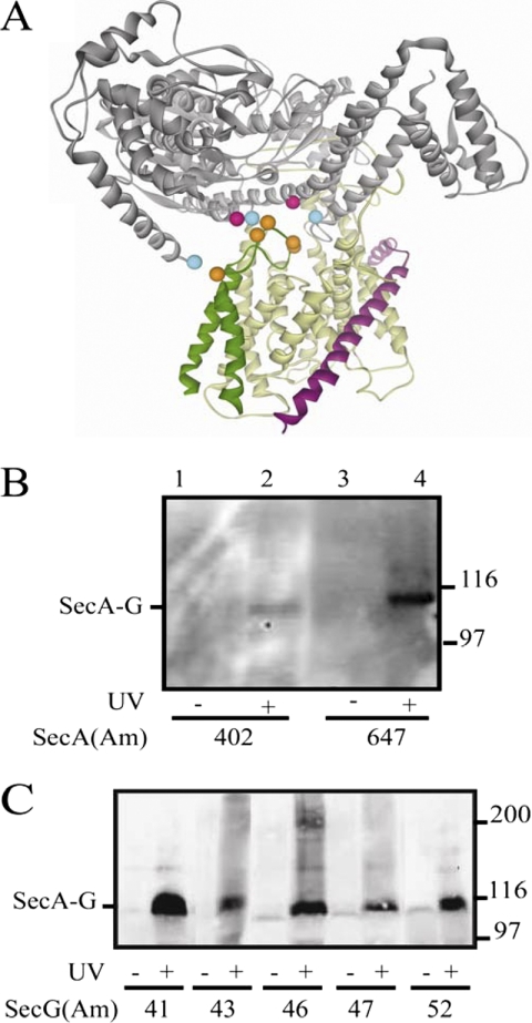
The two major components of the Eubacteria Sec-dependent protein translocation system are the heterotrimeric channel-forming component SecYEG and its binding partner, the SecA ATPase nanomotor. Once bound to SecYEG, the preprotein substrate, and ATP, SecA undergoes ATP-hydrolytic cycles that drive the stepwise translocation of proteins. Although a previous site-directed in vivo photocross-linking study (Mori, H., and Ito, K. (2006) Proc. Natl. Acad. Sci. U.S.A. 103, 16159-16164) elucidated residues of SecY needed for interaction with SecA, no reciprocal study for SecA protein has been reported to date. In the present study we mapped residues of SecA that interact with SecY or SecG utilizing this approach. Our results show that distinct domains of SecA on two halves of the molecule interact with two corresponding SecY partners as well as with the central cytoplasmic domain of SecG. Our data support the in vivo relevance of the Thermotoga maritima SecA·SecYEG crystal structure that visualized SecYEG interaction for only one-half of SecA as well as previous studies indicating that SecA normally binds two molecules of SecYEG.


Figures

FIGURE 1.
FIGURE 1.
Incorporation of pBPA into SecA protein. The secA59(Am) mutant was grown in the presence or absence of pBPA and/or isopropyl-β-d-thiogalactopyranoside (IPTG) as indicated. Cell cultures were grown and analyzed as described under “Experimental Procedures.” Western blotting with Ni+2-conjugated horseradish peroxidase is shown. The position of SecA protein is given.
FIGURE 2.
FIGURE 2.
Specificity of SecA·SecY photocross-linking. The indicated secA and secY plasmid-bearing strain was grown and subjected to photocross-linking (+) or not (−), and cells were analyzed as described in “Experimental Procedures.” Western blotting utilized either SecA antisera (A) or c-Myc antibody (B). The positions of SecA, the SecA·SecY cross-linked complex, and molecular weight markers are given. Only the upper portion of the 20-cm-long gel utilized for better separation of high molecular weight proteins was transferred by electroblotting.
FIGURE 3.
FIGURE 3.
Location of SecY-reactive SecA residues on the T. maritima SecA·SecYEG crystal structure. SecA domains are depicted as colored ribbons as follows: dark blue, NBD-I; cyan, NBD-II; orange, PPXD; green, HSD; light green, HWD. SecYEG is depicted in space filling (A) or ribbons (B) as follows; SecY is in gray or yellow, SecE is in magenta, and SecG is in dark green. A, the SecA·SecYEG complex is viewed from the cytosol (left) or the side (right). SecA residues that were positive or negative for cross-linking to SecY are shown as pink or yellow balls, respectively. The red rectangle outlines the NBD-I-NBD-II half of SecA for which logical SecY acceptors are missing. B, shown is the proximity of SecY-reactive SecA residues within the PPXD-HSD-HWD half of SecA to SecY cytosolic domains. The cytosolic domains of SecY are colored as follows; C2 is in dark red, C4 is in cyan, C5 is in dark blue, and C6 is in pink. SecY-reactive SecA residues are colored according to their most proximal SecY cytosolic domain, which was determined utilizing PyMOL with the appropriate script.
FIGURE 4.
FIGURE 4.
Photocross-linking analysis of secA or secA secY double amber mutant strains. A, the indicated single or double secA(Am) mutant strain was grown and subjected to photocross-linking (+) or not (−), and cells were analyzed by Western blotting utilizing c-Myc antibody as described under “Experimental Procedures.” SecA1-SecY1 and SecA1-SecY2 complexes are indicated. B, shown is a scheme depicting the generation of either (I and II) SecA1-SecY1 or (III) SecA1-SecY2 complexes for the secA59(Am) and secY434(Am) or secA59(Am) secY434(Am) double mutant strains, respectively. N and C designate the two halves of SecA protein comprised of NBD-I-NBD-II or PPXD-HSD-HWD-CTL, respectively, whereas YEG designates each SecYEG protomer. C, the indicated secA(Am) or secY(Am) single or double mutant strain was grown and subjected to photocross-linking (+) or not (−), and cells were analyzed by Western blotting utilizing either SecA antisera (lanes 1–4) or c-Myc antibody (lanes 5–6) as described under “Experimental Procedures.”
FIGURE 5.
FIGURE 5.
Comparison of SecA·SecY photocross-linking results with front-to-front and back-to-back SecA·SecYEG models. Front-to-front (A) and back-to-back (B) models of SecA·SecYEG were generated from the T. maritima SecA·SecYEG crystal structure (38) as described under “Experimental Procedures.” SecA and SecYEG are depicted in colored ribbons as described in the Fig. 3 legend except that the two SecY molecules are shown in light yellow or light gray to better highlight the different orientations. SecA residues that were positive or negative for cross-linking to SecY are shown as pink or yellow balls, respectively. The red rectangle outlines the NBD-I-NBD-II half of SecA, whose SecY-reactive resides are better accommodated by the front-to-front orientation of the SecYEG homodimer.
FIGURE 6.
FIGURE 6.
Photocross-linking analysis of secA and secG amber mutant strains. A, SecA is in gray, SecY is in yellow, SecE is in magenta, and SecG is in dark green. The location on the T. maritima SecA·SecYEG crystal structure (38) of residues tested for photocross-linking is depicted as colored balls; SecA residues 402 and 647 that gave positive results are colored in pink, SecA residues 2, 403, and 800 that gave negative results are colored in cyan, and SecG residues 41, 43, 46, 47, and 52 that all gave positive results are colored in orange. B and C, the indicated secA(Am) (B) or secG(Am) (C) mutant strain was grown and subjected to photocross-linking (+) or not (−), and cells were analyzed by Western blotting utilizing c-Myc antibody as described under “Experimental Procedures.” The positions of the SecA·SecG cross-linked complex and molecular weight markers are given.

References

    1. Osborne A. R., Rapoport T. A., van den Berg B. (2005) Annu. Rev. Cell Dev. Biol. 21, 529–550 - PubMed
    1. Van den Berg B., Clemons W. M., Jr., Collinson I., Modis Y., Hartmann E., Harrison S. C., Rapoport T. A. (2004) Nature 427, 36–44 - PubMed
    1. Mori H., Ito K. (2001) Trends Microbiol. 9, 494–500 - PubMed
    1. Veenendaal A. K., van der Does C., Driessen A. J. (2004) Biochim. Biophys. Acta 1694, 81–95 - PubMed
    1. Papanikou E., Karamanou S., Economou A. (2007) Nat. Rev. Microbiol. 5, 839–851 - PubMed

MeSH terms

LinkOut - more resources