Review
. 2011 Apr;10(4):474-82.
doi: 10.1128/EC.00314-10. Epub 2011 Feb 11.

Mitosis in the human malaria parasite Plasmodium falciparum

Affiliations
Review

Mitosis in the human malaria parasite Plasmodium falciparum

Noel Gerald et al. Eukaryot Cell. 2011 Apr.

Abstract

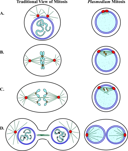
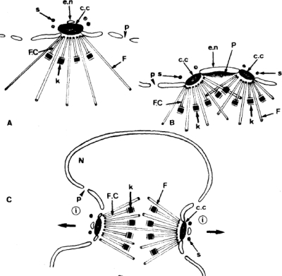
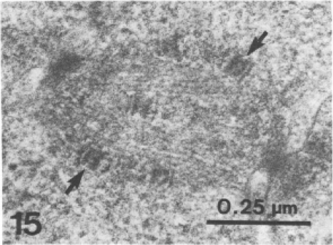
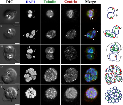
Malaria is caused by intraerythrocytic protozoan parasites belonging to Plasmodium spp. (phylum Apicomplexa) that produce significant morbidity and mortality, mostly in developing countries. Plasmodium parasites have a complex life cycle that includes multiple stages in anopheline mosquito vectors and vertebrate hosts. During the life cycle, the parasites undergo several cycles of extreme population growth within a brief span, and this is critical for their continued transmission and a contributing factor for their pathogenesis in the host. As with other eukaryotes, successful mitosis is an essential requirement for Plasmodium reproduction; however, some aspects of Plasmodium mitosis are quite distinct and not fully understood. In this review, we will discuss the current understanding of the architecture and key events of mitosis in Plasmodium falciparum and related parasites and compare them with the traditional mitotic events described for other eukaryotes.


Figures

Fig. 1.
Fig. 1.
Comparison of traditional views of mitosis with current knowledge of mitosis in blood-stage Plasmodium parasites. A schematic cartoon of mitosis indicating plasma membranes (black lines), microtubule organizing centers (MTOCs, red circles), microtubules (green lines), kinetochores (tan ovals), nuclear membranes (dark blue lines), condensed chromosomes (light blue), and uncondensed chromosomes (light blue with stipple pattern). (A) Early mitosis. In early traditional mitosis (prophase), chromosomes begin to condense within the nuclear membrane, and cytoplasmic microtubules are nucleated by two cytoplasmic MTOCs. In early Plasmodium mitosis, chromosomes remain uncondensed, the two MTOCs are embedded in the nuclear membrane, and microtubules begin to form inside the nucleus. (B) Metaphase. In traditional mitosis, the nuclear membrane has disassembled, condensed chromosomes are attached to the bipolar spindle microtubules through the kinetochores, and the kinetochores are aligned at the metaphase plate. In Plasmodium mitosis, the nuclear membrane remains intact, the chromosomes remain uncondensed, and the kinetochores are captured by the bipolar mitotic spindle forming inside the nucleus. (C) Anaphase. In traditional mitosis, sister chromatids and their associated kinetochores separate and begin to move to opposite poles of the spindle. In Plasmodium, sister kinetochores separate and migrate to opposite spindle poles while the sister chromatids remain uncondensed. (D) Telophase. In traditional views, nuclear membranes assemble around each daughter genome, chromosomes begin to decondense, and the cell begins to divide. In Plasmodium blood-stage schizogony, the nuclear membrane divides to separate daughter genomes and the cell does not divide until several cycles of mitosis have produced a multinuclear cell.
Fig. 2.
Fig. 2.
Interpretation of hemispindle and full mitotic spindle development in Plasmodium from TEM. Images of mitosis during sporogony in P. berghei. (A) A single hemispindle. (B) Hemispindle duplication. (C) Formation of a full mitotic spindle. Microtubules (F and F.C.), MTOCs (c.c.), and kinetochores (k) are indicated. p, nuclear pore; e.n., nuclear envelope; s, cystplasmic satellite; P, bridge of dense material; N, nucleus. (Reprinted from reference with permission of the publisher.)
Fig. 3.
Fig. 3.
TEM image of a mitotic spindle at the beginning of anaphase in a P. falciparum blood-stage parasite. One thin-section image is shown. MTOCs marking the mitotic spindle poles are visible as dense masses at the northwest and southeast corners of the image. Two kinetochores attached to spindle microtubules are indicated by arrows. The remaining kinetochores are visible in other serial sections from this sample. (Reprinted from reference with permission of the publisher.)
Fig. 4.
Fig. 4.
Fluorescence light microscopy images of mitotic spindle during blood-stage schizogony in P. falciparum parasites. 3D confocal microscopy views of blood-stage P. falciparum parasite cell morphology (DIC microscopy), nuclei (DAPI, blue in merged images), mitotic spindle (anti-alpha-tubulin antibody, green in merged images), and mitotic spindle MTOCs (anti-PfCEN3 antibody in rows i, ii, iv, and v or anti-Cr Centrin1 antibody 20H5 in row iii, red in merged images) as previously described (42). Schematic cartoons are drawn for each example, indicating spindle MTOCs (red circles), microtubules (green lines), and outlines of stained DNA (blue lines). Dotted black lines indicate separate parasites in multiply infected host cells. Row i, a and b, short mitotic spindles bounded by MTOCs. The spindle in nucleus b which resembles an oblong tubulin spot has a pole-to-pole distance that is approximately 1 μm long, similar to the lengths of anaphase spindles as described by TEM (52) (also see Fig. 3). Rows ii and iii, c to f, larger spindle structures. In d and e, spindle extends across separate nuclear bodies. In f, a dark line is visible down the center of the DAPI-stained DNA near the spindle midzone (DAPI, arrows). Row iv, asynchronous schizont nuclei have spindles with different geometries in multiple stages of extension. Row v, segmented parasites after the final mitosis of schizogony. Furrows are visible between daughters (arrows), nuclei are condensed, and microtubules appear in the daughter cytoplasms. Row i is a single confocal slice from a 3D series. Rows ii to v are maximum-projection images of full 3D series. Rows ii to iv were processed with deconvolution software. Scale bar, 2 μm.

References

    1. Adisa A., et al. 2007. Re-assessing the locations of components of the classical vesicle-mediated trafficking machinery in transfected Plasmodium falciparum. Int. J. Parasitol. 37:1127–1141 - PubMed
    1. Aikawa M. 1971. Parasitological review. Plasmodium: the fine structure of malarial parasites. Exp. Parasitol. 30:284–320 - PubMed
    1. Aikawa M., Beaudoin R. L. 1968. Studies on nuclear division of a malarial parasite under pyrimethamine treatment. J. Cell Biol. 39:749–754 - PMC - PubMed
    1. Alano P., Billker O. 2005. Gametocytes and gametes, p. 191–219 In Sherman I. W. (ed.), Molecular approaches to malaria. ASM Press, Washington, DC
    1. Aly A. S. I., Vaughan A. M., Kappe S. H. I. 2009. Malaria parasite development in the mosquito and infection of the mammalian host. Annu. Rev. Microbiol. 63:195–221 - PMC - PubMed

Publication types

MeSH terms