. 2011 Jun 1;183(11):1490-8.
doi: 10.1164/rccm.201009-1409OC. Epub 2011 Feb 11.

Leptin promotes fibroproliferative acute respiratory distress syndrome by inhibiting peroxisome proliferator-activated receptor-γ

Affiliations

Leptin promotes fibroproliferative acute respiratory distress syndrome by inhibiting peroxisome proliferator-activated receptor-γ

Manu Jain et al. Am J Respir Crit Care Med. .

Abstract

Rationale: Diabetic patients have a lower incidence of acute respiratory distress syndrome (ARDS), and those who develop ARDS are less likely to die. The mechanisms that underlie this protection are unknown.

Objectives: To determine whether leptin resistance, a feature of diabetes, prevents fibroproliferation after lung injury.

Methods: We examined lung injury and fibroproliferation after the intratracheal instillation of bleomycin in wild-type and leptin-resistant (db/db) diabetic mice. We examined the effect of leptin on transforming growth factor (TGF)-β(1)-mediated transcription in primary normal human lung fibroblasts. Bronchoalveolar lavage fluid (BAL) samples from patients with ARDS and ventilated control subjects were obtained for measurement of leptin and active TGF-β(1) levels.

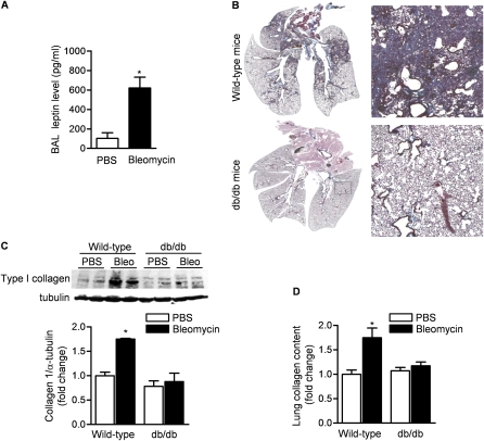
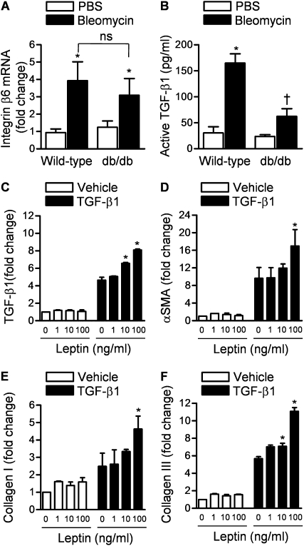
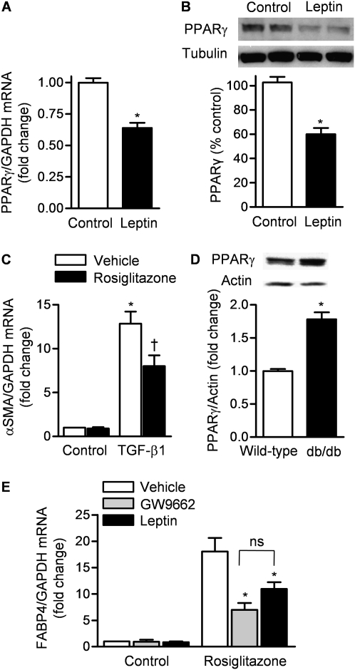
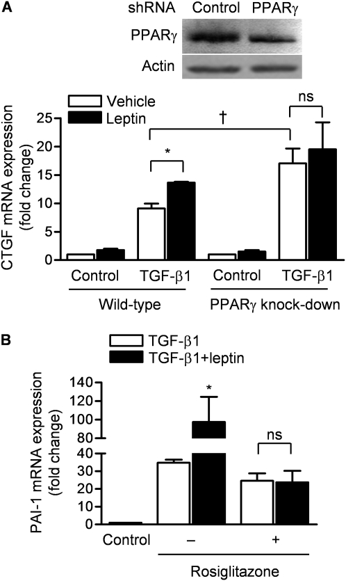
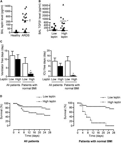
Measurements and main results: Diabetic mice (db/db) were resistant to lung fibrosis. The db/db mice had higher levels of peroxisome proliferator-activated receptor-γ (PPARγ), an inhibitor of the transcriptional response to TGF-β(1), a cytokine critical in the pathogenesis of fibroproliferative ARDS. In normal human lung fibroblasts, leptin augmented the transcription of profibrotic genes in response to TGF-β(1) through a mechanism that required PPARγ. In patients with ARDS, BAL leptin levels were elevated and correlated with TGF-β(1) levels. Overall, there was no significant relationship between BAL leptin levels and clinical outcomes; however, in nonobese patients, higher BAL leptin levels were associated with fewer intensive care unit- and ventilator-free days and higher mortality.

Conclusions: Leptin signaling is required for bleomycin-induced lung fibrosis. Leptin augments TGF-β(1) signaling in lung fibroblasts by inhibiting PPARγ. These findings provide a mechanism for the observed protection against ARDS observed in diabetic patients.


Figures

Figure 1.
Figure 1.
Mice with leptin resistance are protected against bleomycin-induced pulmonary fibrosis. (A) Bronchoalveolar lavage (BAL) fluid levels of leptin in wild-type mice 5 days after intratracheal instillation of bleomycin (0.075 unit) or phosphate-buffered saline (PBS). (B) Masson's trichrome staining for collagen in lungs from mice (wild-type and db/db) 21 days after intratracheal instillation of bleomycin or PBS. Both low-power images were captured with Neurolucida software (MBF Bioscience, Williston, VT) (original magnification: ×5) and high-power field views (original magnification, ×200) are shown. Total collagen content in lungs at 21 days after bleomycin treatment was assessed by (C) collagen I immunoblotting and (D) picrosirius red collagen precipitation (*P < 0.05, bleomycin vs. PBS treatment; n = 8 in each treatment group from two independent experiments).
Figure 2.
Figure 2.
Protection against bleomycin-induced pulmonary fibrosis is independent of bleomycin-induced lung injury in mice. (A) Hematoxylin and eosin–stained lungs from mice (wild-type and db/db) 5 days after the intratracheal administration of bleomycin. Both low-power images were captured with MBF Neurolucida (original magnification: ×5) and high-power field views (original magnification: ×200) are shown. (B) Cell count and (C) levels of proinflammatory cytokines/chemokines in the bronchoalveolar lavage (BAL) fluid from mice (wild-type and db/db) 5 days after the intratracheal administration of bleomycin. (*P < 0.05, bleomycin vs. phosphate-buffered saline [PBS] treatment; n = 5 in each treatment group). MCP-1 = monocyte chemotactic protein-1; TNF-α = tumor necrosis factor-α; ns = not significant.
Figure 3.
Figure 3.
Leptin signaling affects bleomycin-induced transforming growth factor (TGF)-β1 activation downstream of up-regulation of integrin αvβ6. (A) Real-time quantitative mRNA levels of integrin αvβ6 (corrected to keratin mRNA) (*P < 0.05, bleomycin vs. phosphate-buffered saline [PBS]; ns = not significant). (B) Bronchoalveolar lavage (BAL) fluid levels of TGF-β1 in wild-type and db/db mice 5 days after intratracheal instillation of bleomycin or PBS (P < 0.05: *bleomycin vs. PBS, db/db plus bleomycin vs. wild-type plus bleomycin). (C) Normal human lung fibroblasts were treated with either vehicle or TGF-β1 (5 ng/ml) in the presence of various concentrations of leptin and TGF-β1 mRNA was measured 24 hours later by quantitative real-time reverse transcription PCR (qRT-PCR). (DF) Normal human lung fibroblasts were treated with either vehicle or TGF-β1 (5 ng/ml) in the presence of various concentrations of leptin, and α-smooth muscle actin (α-SMA), collagen I, and collagen III mRNAs were measured 24 hours later (qRT-PCR). (*P < 0.05 leptin vs. PBS) (n ≥ 4 in each treatment group from three independent experiments).
Figure 4.
Figure 4.
Leptin decreases the expression and activity of the transforming growth factor (TGF)-β1 suppressor, peroxisome proliferator–activated receptor-γ (PPARγ). (A) mRNA (quantitative real-time reverse transcription PCR [qRT-PCR]) and (B) protein (immunoblot) levels of PPARγ in normal human lung fibroblasts 24 hours after treatment with leptin (100 ng/ml) or vehicle (*P < 0.05, leptin vs. control treatment). GAPDH = glyceraldehyde-3-phosphate dehydrogenase. (C) α-Smooth muscle actin (α-SMA) mRNA (qRT-PCR) in normal human lung fibroblasts 24 hours after treatment with TGF-β1 (5 ng/ml) or vehicle (control) in the presence or absence of rosiglitazone (50 μM) (a PPARγ agonist) (P < 0.05: *TGF-β1 vs. control, TGF-β1 plus vehicle vs. TGF-β1 plus rosiglitazone). (D) PPARγ protein levels (immunoblot) in mouse lungs from untreated wild-type and db/db mice (P < 0.05: *wild-type vs. db/db). (E) mRNA (qRT-PCR) for fatty acid–binding protein-4 (FABP4) in normal human lung fibroblasts (NHLFs) treated with rosiglitazone (50 μM) in the presence or absence of GW9662 (a PPARγ antagonist) (10 μM) and leptin (100 ng/ml) (P < 0.05: *GW9662 vs. vehicle, *leptin vs. Vehicle; ns = not significant) (n ≥ 4 in each treatment group from three independent experiments).
Figure 5.
Figure 5.
Leptin-mediated augmentation of transforming growth factor (TGF)-β1 transcription in lung fibroblasts requires peroxisome proliferator–activated receptor-γ (PPARγ). (A) Normal human lung fibroblasts (NHLFs) were stably transfected (lentivirus) with control short hairpin RNA (shRNA) or an shRNA against PPARγ and cell lysates were immunoblotted for PPARγ (top). These cells were treated with medium and TGF-β1 (5 ng/ml) in the absence or presence of leptin (100 ng/ml) and 24 hours later connective tissue growth factor (CTGF) mRNA expression was measured (quantitative real-time reverse transcription PCR [qRT-PCR]) (P < 0.05: *leptin vs. vehicle, wild-type plus TGF-β1 plus vehicle vs. PPARγ knockdown plus TGF-β1 plus vehicle; ns = not significant). (B) NHLFs were treated with TGF-β1 (5 ng/ml), leptin (100 ng/ml), and/or rosiglitazone (50 μM) and 24 hours later plasminogen activator inhibitor (PAI)-1 mRNA was measured (qRT-PCR) (*P < 0.05, TGF-β1 vs. TGF-β1 plus leptin) (n = 4 in each treatment group from two independent experiments).
Figure 6.
Figure 6.
Alveolar levels of leptin and transforming growth factor (TGF)-β1 correlate in patients with acute respiratory distress syndrome (ARDS). (A) Bronchoalveolar lavage (BAL) fluid levels of leptin in healthy intubated control patients and all patients with ARDS. (B) BAL fluid levels of TGF-β1 (all patients) and (C) clinical outcomes (ventilator-free days, intensive care unit [ICU]–free days) and (D) survival of patients with ARDS with low (<100 pg/ml) and high (>100 pg/ml) levels of leptin. (P < 0.05: ARDS vs. healthy control subjects, *high leptin vs. low leptin) (n = 36 patients with ARDS and n = 15 healthy intubated patients). BMI = body mass index.

References

    1. Rubenfeld GD, Caldwell E, Peabody E, Weaver J, Martin DP, Neff M, Stern EJ, Hudson LD. Incidence and outcomes of acute lung injury. N Engl J Med 2005;353:1685–1693. - PubMed
    1. Moss M, Guidot DM, Steinberg KP, Duhon GF, Treece P, Wolken R, Hudson LD, Parsons PE. Diabetic patients have a decreased incidence of acute respiratory distress syndrome. Crit Care Med 2000;28:2187–2192. - PubMed
    1. Frank JA, Nuckton TJ, Matthay MA. Diabetes mellitus: a negative predictor for the development of acute respiratory distress syndrome from septic shock. Crit Care Med 2000;28:2645–2646. - PubMed
    1. Gajic O, Dabbagh O, Park PK, Adesanya A, Chang SY, Hou P, Anderson H III, Hoth JJ, Mikkelsen ME, Gentile NT, et al.; on behalf of the U.S. Critical Illness and Injury Trials Group: Lung Injury Prevention Study Investigators. Early identification of patients at risk of acute lung injury: evaluation of lung injury prediction score in a multicenter cohort study. Am J Respir Crit Care Med 2010;183:462–470. - PMC - PubMed
    1. Gong MN, Thompson BT, Williams P, Pothier L, Boyce PD, Christiani DC. Clinical predictors of and mortality in acute respiratory distress syndrome: potential role of red cell transfusion. Crit Care Med 2005;33:1191–1198. - PubMed

Publication types