. 2011 Mar 1;108(9):3473-80.
doi: 10.1073/pnas.1017075108. Epub 2011 Feb 11.

Development of an artificial cell, from self-organization to computation and self-reproduction

Affiliations

Development of an artificial cell, from self-organization to computation and self-reproduction

Vincent Noireaux et al. Proc Natl Acad Sci U S A. .

Abstract

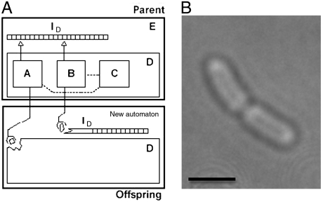
This article describes the state and the development of an artificial cell project. We discuss the experimental constraints to synthesize the most elementary cell-sized compartment that can self-reproduce using synthetic genetic information. The original idea was to program a phospholipid vesicle with DNA. Based on this idea, it was shown that in vitro gene expression could be carried out inside cell-sized synthetic vesicles. It was also shown that a couple of genes could be expressed for a few days inside the vesicles once the exchanges of nutrients with the outside environment were adequately introduced. The development of a cell-free transcription/translation toolbox allows the expression of a large number of genes with multiple transcription factors. As a result, the development of a synthetic DNA program is becoming one of the main hurdles. We discuss the various possibilities to enrich and to replicate this program. Defining a program for self-reproduction remains a difficult question as nongenetic processes, such as molecular self-organization, play an essential and complementary role. The synthesis of a stable compartment with an active interface, one of the critical bottlenecks in the synthesis of artificial cell, depends on the properties of phospholipid membranes. The problem of a self-replicating artificial cell is a long-lasting goal that might imply evolution experiments.


Conflict of interest statement

The authors declare no conflict of interest.

Figures

Fig. 1.
Fig. 1.
Cells observed by Robert Hooke and Theodor Schwann. (Top) Cells observed through a slice of cork with a microscope in 1665 by Robert Hooke (8). Hooke saw a pattern of holes that he named cells (each cell about 20 μm). In the figure on the left, the cells are split the “long ways” and show “diaphragms” between cells as described by Hooke. A branch of the cork oak tree is shown below. Reprinted from ref. . (Bottom: images 1 to 14) Plant and animal cells observed by Theodor Schwann in 1839 (10) (figures 1, 2, 3, and 14 are from plants, figures 4, 5, 6, and 7 are from a fish, and figures 10, 11, and 12 are from an animal eye). Reprinted from ref. .
Fig. 2.
Fig. 2.
A self-reproducing automaton and a bacterium. (A) Von Neumann’s logic. A universal constructor (part A) constructs the hardware (part D) of the offspring automaton according to the instruction I. The copier B makes a copy of the software part of the offspring automaton, the instruction tape I. Part A and part B are controlled by the regulatory part C. (B) Phase contrast microscopy image of the division of an E. coli cell (scale bar: 1 μm).
Fig. 3.
Fig. 3.
Example of nongenetic processes. (A) Cross-sections of the three structures that self-organize upon the dispersion of phospholipids in aqueous solutions. Phospholipids molecules are composed of a hydrophilic head (red circles) and of two hydrophobic chains (dark region). The interior of spherical micelles is composed of hydrophobic chains only. Bilayer structures arranged in spherical shapes with aqueous solutions inside and outside are called liposomes or vesicles. Large planar bilayers can also be formed. (B) A picture illustrating molecular crowding in the cytoplasm of E. coli. In green: the phospholipid membrane, the cell wall, and a flagellum. In purple: the ribosomes synthesizing proteins (white). In blue: the high protein concentration in the cytoplasm. In yellow: DNA with interacting DNA binding proteins [Reproduced with permission from Dr. David S. Goodsell (Copyright 2000, David S. Goodsell).] (C) α-hemolysin pore formation, an example of protein self-organization at the membrane. The soluble monomer (step 1) interacts with the bilayer (step 2) to form a prepore composed of seven monomers (step 3) before pore formation (step 4) through the membrane [Reproduced with permission from ref.  (Copyright 2003, Elsevier).]
Fig. 4.
Fig. 4.
Protein expression in a test tube, in a vesicle, and in a vesicle with pores. (A) Kinetics of expression of eGFP in a test tube (open circles), in a phospholipid vesicle (closed dark circles), and expression of α-hemolysin-eGFP in a phospholipid vesicle (closed green circles). (Inset) Blowup of the first 20 h. (B) Fluorescence microscopy images of α-hemolysin-eGFP expressed inside vesicles after a few hours of expression [(Top to Bottom) a vesicle doublet, a single vesicle, and an aggregate of vesicles]. Scale bar: 10 μm. [Reproduced with permission from ref.  (Copyright 2004, National Academy of Sciences).]
Fig. 5.
Fig. 5.
Self-organization of polymeric structures inside phospholipid vesicles. (A) Filamentous actin (F-actin) with fascin in a liposome. Scale bar 5 μm. [Reproduced with permission from ref.  (Copyright 1999, Elsevier).] (B) F-actin and anchoring proteins (ankyrin and spectrin) in a liposome. Red: lipid, Green: F-actin. Scale bar 5 μm. [Reproduced with permission from ref.  (Copyright Wiley-VCH Verlag GmbH & Co.).] (C) Microtubule in a liposome. Buckling of microtubule using micropipette aspiration (the arrow show the position of the vesicle in the pipette). Microtubule is shown as red line, whereas membrane is a black line in the schematic illustration. [Reproduced with permission from ref.  (Copyright 1996, American Physical Society).] (D) Cell-free expression of the bacterial actin MreB and its associating membrane protein MreC inside a liposome. Green: YFP-MreB, Red: Rhodamine-BSA (used as a fluorescent cytoplasmic marker). YFP-MreB form filaments as it interacts with MreC. Scale bar: 10 μm.

References

    1. Schrödinger E. What Is Life? The Physical Aspect of the Living Cell. Cambridge, England: University Press; 1945. p. viii.
    1. Crick F. Life Itself: Its Origin and Nature. New York: Simon & Schuster; 1981.
    1. Lane N. Power, Sex, Suicide: Mitochondria and the Meaning Of Life. Oxford, New York: Oxford Univ Press; 2005. p. xiii.
    1. Von Neumann J. The general and logical theory of automata. In: Jeffress LA, editor. Cerebral Mechanisms in Behavior: The Hixon Symposium. New York: Wiley; 1951.
    1. Kauffman SA. At Home in the Universe: The Search for Laws of Self-Organization and Complexity. New York: Oxford Univ Press; 1995. p. viii.

Publication types

MeSH terms