Comparative Study
. 2011 Mar 15;186(6):3674-85.
doi: 10.4049/jimmunol.1003037. Epub 2011 Feb 11.

Supplemental and highly elevated tocopherol doses differentially regulate allergic inflammation: reversibility of α-tocopherol and γ-tocopherol's effects

Affiliations
Comparative Study

Supplemental and highly elevated tocopherol doses differentially regulate allergic inflammation: reversibility of α-tocopherol and γ-tocopherol's effects

Christine A McCary et al. J Immunol. .

Abstract

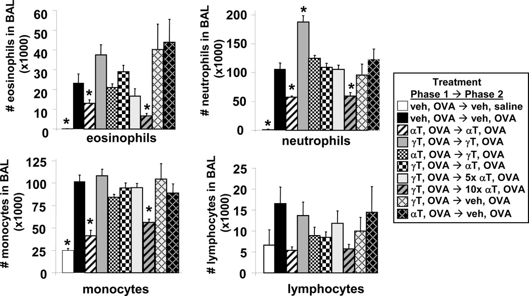
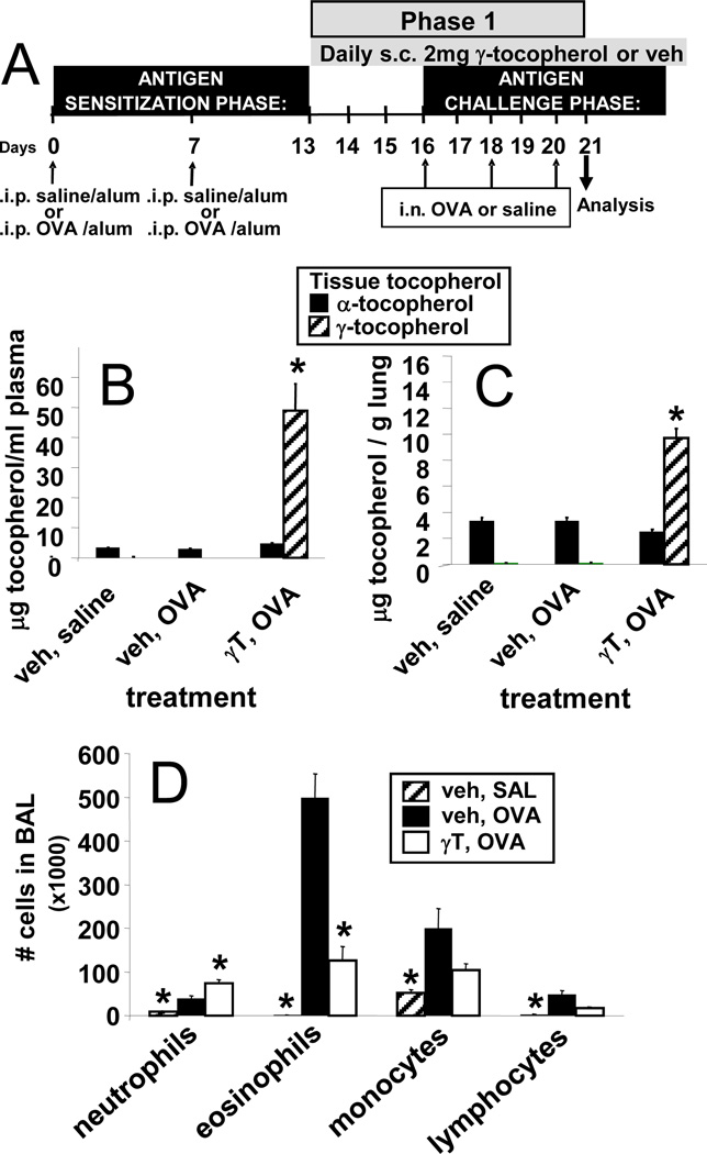
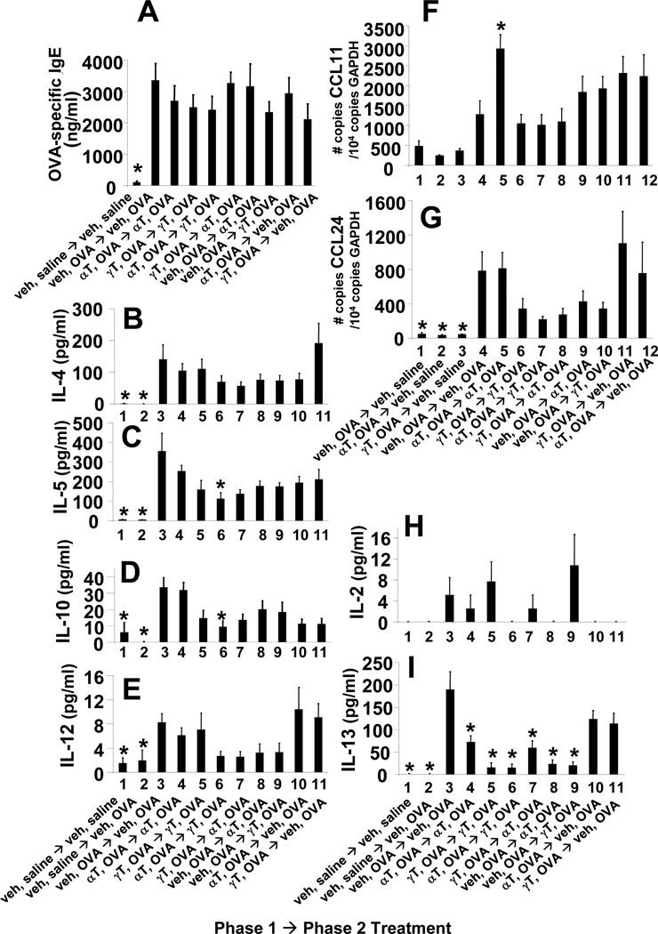
We have reported that supplemental doses of the α- and γ-tocopherol isoforms of vitamin E decrease and increase, respectively, allergic lung inflammation. We have now assessed whether these effects of tocopherols are reversible. For these studies, mice were treated with Ag and supplemental tocopherols in a first phase of treatment followed by a 4-wk clearance phase, and then the mice received a second phase of Ag and tocopherol treatments. The proinflammatory effects of supplemental levels of γ-tocopherol in phase 1 were only partially reversed by supplemental α-tocopherol in phase 2, but were completely reversed by raising α-tocopherol levels 10-fold in phase 2. When γ-tocopherol levels were increased 10-fold (highly elevated tocopherol) so that the lung tissue γ-tocopherol levels were equal to the lung tissue levels of supplemental α-tocopherol, γ-tocopherol reduced leukocyte numbers in the lung lavage fluid. In contrast to the lung lavage fluid, highly elevated levels of γ-tocopherol increased inflammation in the lung tissue. These regulatory effects of highly elevated tocopherols on tissue inflammation and lung lavage fluid were reversible in a second phase of Ag challenge without tocopherols. In summary, the proinflammatory effects of supplemental γ-tocopherol on lung inflammation were partially reversed by supplemental levels of α-tocopherol but were completely reversed by highly elevated levels of α-tocopherol. Also, highly elevated levels of γ-tocopherol were inhibitory and reversible in lung lavage but, importantly, were proinflammatory in lung tissue sections. These results have implications for future studies with tocopherols and provide a new context in which to review vitamin E studies in the literature.


Figures

Figure 1
Figure 1. Timeline synopsis for tocopherol and OVA treatments
A) Tocopherol treatment during a single phase of OVA challenges. B) Tocopherol treatment during two phases of OVA challenges.
Figure 2
Figure 2. Opposing functions of supplemental levels of natural d-α-tocopherol and d-γ-tocopherol
A. Structure of natural d-α-tocopherol (d-α-T) and natural d-γ-tocopherol (d-γ-T), which differ by one methyl group (arrows). B. Tocopherol in plasma and perfused lung tissue after 8 daily subcutaneous (s.c.) doses of 0.04, 0.2 or 0.6 mg αT or γT. Tocopherol was measured by HPLC. n = 4–5 animals per group. C. Allergic lung inflammation protocol with intraperitoneal (i.p.) sensitization with chicken egg white albumin (OVA) fraction V and tocopherol treatment starting after OVA-sensitization. Intranasal (i.n.) OVA challenge was with fraction VI OVA. Inflammation was analyzed 24 hours after the last OVA challenge. D. Bronchoalveolar lavage (BAL) neutrophils, eosinophils, monocytes and lymphocytes were counted with a hemocytometer and cytospun BAL cells were counted by standard morphological criteria. Veh, vehicle.
Figure 3
Figure 3. Timeline and plasma tocopherol levels during analysis of the reversibility of the regulatory effects of supplemental tocopherol on allergic lung inflammation
A. Mice were sensitized with OVA and then administered subcutaneous 0.2 mg tocopherol daily during OVA challenge in phase 1 of treatments. Following phase 1, mice entered a 4-week clearance phase during which they were not treated with tocopherols or challenged with OVA. At the end of the clearance phase, mice were treated subcutaneously with 0.2 mg tocopherols and rechallenged with OVA in phase 2 of treatments. In phase 2, mice were again treated with daily doses of supplemental tocopherol (same isoform as phase 1, different isoform than phase 1, or vehicle) and re-challenged one time with OVA. In addition, two treatment groups were daily administered 5× and 10× supplemental αT in phase 2. Inflammation was analyzed 24 hours following this re-challenge. n, number of animals per group. B. Tocopherol in plasma on day 21 as measured by HPLC. C. Tocopherol in plasma on day 49 as measured by HPLC. D. Tocopherol in plasma on day 54 as measured by HPLC. Note that on day 21, day 49 or day 54, plasma tocopherol in saline-treated groups receiving tocopherols were the same as those treated with OVA and tocopherols (data not shown). i.p., intraperitoneal; i.n., intranasal; veh, vehicle; αT, d-alpha-tocopherol; γT, d-gamma-tocopherol; veh OVA→veh OVA group, indicates phase 1 treatments followed by phase 2 treatments as in panel A.
Figure 4
Figure 4. Reversibility of the regulatory effects of supplemental tocopherol levels on allergic bronchoalveolar lavage inflammation
Mice were treated with tocopherols and OVA as in Figure 3. On day 54, bronchoalveolar lavage (BAL) neutrophils, eosinophils, monocytes and lymphocytes were counted with a hemocytometer and cytospun BAL cells were counted by standard morphological criteria. veh OVA→veh OVA group, indicates phase 1 treatments followed by phase 2 treatments as described in Figure 3A; αT, d-alpha-tocopherol; γT, d-gamma-tocopherol; *, p<0.05 as compared to veh OVA→veh OVA group.
Figure 5
Figure 5. Reversibility of the regulatory effects of supplemental tocopherol on allergic lung tissue inflammation
Mice were treated with tocopherols as in Figure 3A. A–I. Representative micrographs (40× objective) of perivascular regions in the lung tissue from day 54 as in Figure 3A. Lung tissues were stained with hematoxylin and eosin. J. number of blood eosinophils on day 54 of treatments as in Figure 3A. white arrow, vessel lumen; black arrow, bronchial airway; veh OVA→veh OVA group, indicates phase 1 treatments followed by phase 2 treatments as described in Figure 3A; veh, vehicle; αT, d-alpha-tocopherol; γT, d-gamma-tocopherol.
Figure 6
Figure 6. Switching tocopherol isoforms at supplemental levels in phase 2 did not alter OVA-specific IgE, lung cytokines or lung chemokines
Mice were treated with supplemental levels of tocopherols as in Figure 3A. A. serum OVA-specific IgE as measured by ELISA. B-F,I,J. BAL supernatants were examined for cytokines using the Th1/Th2 mouse cytokine multiplexing kit with IL-13 (Life Technologies). G,H. lung tissue was placed in RNAlater then examined for eotaxin 1 (CCL11) and eotaxin 2 (CCL24) expression by real-time PCR. veh OVA→veh OVA group, indicates phase 1 treatments followed by phase 2 treatments as described in Figure 3A; veh, vehicle; αT, d-alpha-tocopherol; γT, d-gamma-tocopherol; *, p<0.05 as compared to veh OVA→veh OVA group.
Figure 7
Figure 7. Highly-elevated γ-tocopherol decreases lung bronchoalveolar lavage inflammation
A. Timeline for treatments with OVA and highly-elevated γ-tocopherol (γT) during allergic lung inflammation (only 1 phase of OVA treatments). On day 21 (panel A), plasma was collected and lungs were perfused free of blood. Tocopherol was measured by HPLC. Lung lavage leukocytes were examined on day 21. B. Plasma tocopherol. C. Lung tocopherols. D. BAL neutrophils, eosinophils, monocytes and lymphocytes were counted with a hemocytometer and cytospun BAL cells were counted by standard morphological criteria. γT, d-gamma-tocopherol; veh, vehicle; *, p<0.05 as compared to veh, OVA group. n = 4 mice per group for the saline group and 8 mice per group for the OVA-stimulated groups.
Figure 8
Figure 8. Highly-elevated γ-tocopherol treatment decreases Th2 cytokines and chemokines in the lung
Mice were treated with highly-elevated γ-tocopherol as in Figure 7A. BAL supernatants were examined for cytokines (IL-4, IL-5, IL-10, IFNγ, IL-2, MIP-1α, MCP-1) using a mouse cytokine multiplexing assay (Millipore). Lung tissue was placed in RNAlater and then examined for eotaxin 1 (CCL11) and eotaxin 2 (CCL24) expression by quantitative real-time PCR. γT, d-gamma-tocopherol; veh, vehicle; *, p<0.05 as compared to veh, OVA group.
Figure 9
Figure 9. Highly-elevated γ-tocopherol reduces leukocyte transendothelial migration through direct regulation of endothelial cells
A. 90% confluent endothelial cell monolayers were treated overnight with a dose curve of γ-tocopherol. After washing, cells were harvested and tocopherol was measured by HPLC. B. 90% confluent endothelial cell monolayers were treated overnight with 20 µM γ-tocopherol. Cells were washed 5 times before the start of the leukocyte migration assay with physiological laminar flow as detailed in Methods. C. Mice were treated with highly-elevated (2 mg/day) γ-tocopherol or vehicle (ethoxylated castor oil) for 4 days as we previously reported for in vivo loading of tocopherols in leukocytes (8). Spleens were collected and red blood cells were lysed by hypotonic lysis. Spleen leukocytes from vehicle mice or spleen leukocytes from mice treated with highly-elevated γ-tocopherol were added to untreated endothelial cell monolayers and examined for transendothelial migration under physiological laminar flow conditions as detailed in methods. Leukocytes in each treatment group were not pooled; leukocytes from each mouse were processed individually. γT, d-gamma-tocopherol; veh, vehicle; *, p<0.05 as compared to vehicle-treated group. n = 3 migration assays per treatment.
Figure 10
Figure 10. Timeline and plasma tocopherol levels during analysis of the reversibility of the regulatory effects of highly-elevated tocopherol levels on allergic lung inflammation
A. Mice were sensitized with OVA and then administered subcutaneous highly-elevated (2 mg) tocopherol daily during OVA challenge in phase 1 of treatments. Following phase 1, mice entered a 4-week clearance phase during which they were not treated with tocopherols or challenged with OVA. At the end of the clearance phase, mice were treated subcutaneously with highly-elevated (2 mg) tocopherols and rechallenged with OVA in phase 2 of treatments. In phase 2, mice were again treated with daily doses of supplemental tocopherol (same isoform as phase 1, different isoform than phase 1, or vehicle) and re-challenged three times with OVA. All treatment groups are listed. B. Plasma tocopherol on day 21. C. Plasma tocopherol on day 49. D. Plasma tocopherol on day 58. E. Tocopherol in perfused lung on day 58. veh OVA→veh OVA group, indicates phase 1 treatments followed by phase 2 treatments as described in Figure 10A; veh, vehicle; αT, d-alpha-tocopherol; γT, d-gamma-tocopherol; *, p<0.05 as compared to veh OVA on days 21 or 49 or as compared to veh OVA→veh OVA group on day 58 where indicated. n, number of mice per group.
Figure 11
Figure 11. Reversibility of the regulatory effects of highly-elevated tocopherol levels on allergic bronchoalveolar lavage inflammation
Mice were treated with highly-elevated tocopherols as in Figure 10A. On day 58, bronchoalveolar lavage (BAL) neutrophils, eosinophils, monocytes and lymphocytes were counted with a hemocytometer. Cytospun BAL cells were counted by standard morphological criteria. veh, vehicle; αT, d-alpha-tocopherol; γT, d-gamma-tocopherol; *, p<0.05 as compared to veh OVA→veh OVA group on day 58. Not shown are two saline groups: leukocytes in the BAL of the αT,OVA→veh,saline and γT,OVA→veh,saline groups had BAL cell infiltrate not significantly different than veh,saline→veh,saline group.
Figure 12
Figure 12. Reversibility of the regulatory effects of highly-elevated tocopherol on allergic lung tissue inflammation
Mice were treated with highly-elevated tocopherols as in Figure 10A. A–E. Representative micrographs (20× objective) of perivascular regions in the lung tissue from day 58 as in Figure 10A. Lung tissue was stained with hematoxylin and eosin. F. number of blood eosinophils on day 58 of treatments as in Figure 10A. white arrow, vessel lumen; black arrow, bronchial airway; veh OVA→veh OVA group, indicates phase 1 treatments followed by phase 2 treatments as described in Figure 10A; veh, vehicle; αT, d-alpha-tocopherol; γT, d-gamma-tocopherol; *, p<0.05 as compared to veh OVA→veh OVA group.
Figure 13
Figure 13. Switching tocopherol isoforms at highly-elevated levels in phase 2 decreased IL-13 but not other lung cytokines, lung chemokines or OVA-specific IgE
Mice were treated with highly-elevated levels of tocopherols as in Figure 10A. A. serum OVA-specific IgE was measured by ELISA. B–E, H–I. BAL supernatants were examined for cytokines using the Th1/Th2 mouse cytokine multiplexing kit plus IL-13 singleplex supplement (Life Technologies). F,G. Lung tissue was placed in RNAlater then examined for eotaxin 1 (CCL11) and eotaxin 2 (CCL24) expression by quantitative real-time PCR. veh OVA→veh OVA group, indicates phase 1 treatments followed by phase 2 treatments as described in Figure 10A; veh, vehicle; αT, d-alpha-tocopherol; γT, d-gamma-tocopherol; *, p<0.05 as compared to veh OVA→veh OVA group.

References

    1. Zingg JM, Azzi A. Non-antioxidant activities of vitamin E. Curr Med Chem. 2004;11:1113–1133. - PubMed
    1. Brigelius-Flohe R, Traber MG. Vitamin E: function and metabolism. FASEB J. 1999;13:1145–1155. - PubMed
    1. Wolf G. How an increased intake of alpha-tocopherol can suppress the bioavailability of gamma-tocopherol. Nutr Rev. 2006;64:295–299. - PubMed
    1. Desrumaux C, Deckert V, Athias A, Masson D, Lizard G, Palleau V, Gambert P, Lagrost L. Plasma phospholipid transfer protein prevents vascular endothelium dysfunction by delivering alpha-tocopherol to endothelial cells. FASEB J. 1999;13:883–892. - PubMed
    1. Atkinson J, Epand RF, Epand RM. Tocopherols and tocotrienols in membranes: a critical review. Free Radic Biol Med. 2008;44:739–764. - PubMed

Publication types

MeSH terms