. 2011 Mar 7;56(5):1397-414.
doi: 10.1088/0031-9155/56/5/012. Epub 2011 Feb 11.

Iterative deconvolution of simultaneous 99mTc and 201Tl projection data measured on a CdZnTe-based cardiac SPECT scanner

Affiliations

Iterative deconvolution of simultaneous 99mTc and 201Tl projection data measured on a CdZnTe-based cardiac SPECT scanner

Krzysztof Kacperski et al. Phys Med Biol. .

Abstract

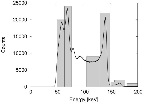
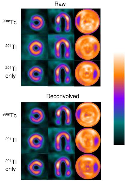
We present a method of correcting self-scatter and crosstalk effects in simultaneous technetium-99m/thallium-201 stress/rest myocardial perfusion (single photon emission computed tomography) SPECT scans. The method, which is in essence a hybrid between the triple energy window method and scatter modelling, is based on a model of spatial and spectral distribution of projection counts in several selected energy windows. The parameters of the model are determined from measurements of thin rod sources in air when no in-object scatter or attenuation effects are present. The model equations are solved using the iterative maximum likelihood expectation maximization algorithm in the projection space to find estimates of the primary photopeak counts of both radionuclides. The method has been developed particularly for a novel dedicated cardiac camera based on CdZnTe pixellated detectors, although it can also be adapted to a conventional scintillator camera. The method has been validated in anthropomorphic phantom experiments. Significant improvement in defect contrast has been observed with only moderate increase in image noise. The application of the method to patient data is illustrated.


Figures

Figure 1
Figure 1
Spectra of uniformly scanned thin rod 99mTc and 201Tl sources measured in air for D-SPECT and conventional gamma camera (Prism 3000, Philips, Cleveland, OH)
Figure 2
Figure 2
Count profiles through the static projections of a 201Tl rod source in air on a single detector head measured in the main photopeak window 64-77 keV and the lower window 50-64 keV containing mainly tungsten fluorescence X-rays. Solid lines represent Gaussians fitted to measured data points.
Figure 3
Figure 3
The width (FWHM) of 201Tl rod source static projections as a function of distance to the detector for the specified energy windows.
Figure 4
Figure 4
Total spectrum of an anthropomorphic phantom filled with 99mTc and 201Tl activities as described in section 2.7 (configuration 1), measured on D-SPECT. The energy windows described in sec. 2.4 are marked.
Figure 5
Figure 5
Simulated distribution of scattered photons for a point 99mTc source in a cylinder 60×60 cm at varying depths. Dotted lines - scatter in the 130-149 keV photopeak window, solid lines - true scatter in the 105-130 keV window, dotted-dashed lines - estimations of scatter in the 105-130 keV window using Eq. 3 with fixed patrameters α3 = 0.43 and λ3 = 0.18 cm−1.
Figure 6
Figure 6
Simulated distribution of scattered photons for a point 201Tl source in a cylinder 60×60 cm at varying depths. Dotted lines - scatter in the 64-77 keV photopeak window, solid lines - true scatter in the 50-64 keV window, dotted-dashed lines - estimations of scatter in the 50-64 keV window using Eq. 3 with fixed patrameters α1 = 0.22 and λ1 = 0.2 cm−1.
Figure 7
Figure 7
A profile through heart projection of scattered counts in the scatter energy window (105-130 keV) generated from the NCAT phantom using Monte Carlo simulation (points) and an approximated scatter distribution obtained by convolving the scatter projection in photopeak window (130-149 keV) with the kernel given by Eq. (3) with α3 = 0.43 and λ3 = 0.18cm−1 (solid line).
Figure 8
Figure 8
Reconstructed images of the phantom with small 100% defects (6 minute scan): short axis slices, horizontal long axis slices and polar map for reconstructions based on raw (windows 2+6 and 4) and deconvolved projections; inter-iterative smoothing with a Gaussian kernel with σ = 1; 99mTc images - 5 iterations, 201Tl images - 10 iterations.
Figure 9
Figure 9
Contrast noise curves for the phantom with large 70% defects. Top graph shows measured true defect contrast (in perfect image it should be 0.7); bottom graph shows contrast in defect regions for non reduced activity (should be 0 in perfect image). Points on each curve correspond to 1, 2, 3, 5, and 10 iterations. Larger false defect contrast in 99mTc images is mainly due to attenuation effect in the inferior wall.
Figure 10
Figure 10
Polar plots of LV 201Tl images of the phantom with large 70% defects (4 minute dual radionuclide scan) reconstructed from raw data ([64 - 77 keV] + [157 - 177 keV] windows), and projections deconvolved with inter-iterative smoothing (Gaussian kernel with σ = 1). Images obtained from 201Tl-only scan of the same phantom aer shown for comparison. Activity concentrations (perfusion) calculated by the QPS software.
Figure 11
Figure 11
Simultaneous 201Tl rest - 99mTc stress study of a 73y old male: (a) uncorrected data reconstruction for 20% 140 keV and 15% 70 keV energy windows; (b) reconstruction from data processed by the iterative deconvolution algorithm; (c) rest image obtaied from uncorrected 201Tl only scan (prior to injection of 99mTc ). The arrows indicate the defect in infero-lateral wall, enhanced after crosstalk/scatter correction.

References

    1. Beekman FJ, de Jong HW, van Geloven S. Efficient fully 3-D iterative SPECT reconstruction with Monte Carlo-based scatter compensation. IEEE Trans Med Imaging. 2002;21(8):867–877. - PubMed
    1. Ben-Haim S, Kacperski K, Hain S, Van Gramberg D, Hutton BF, Erlandsson K, Sharir T, Roth N, Waddington WA, Berman DS, Ell PJ. Simultaneous dual-radionuclide myocardial perfusion imaging with a solid-state dedicated cardiac camera. Eur J Nucl Med Mol Imaging. 2010;37(9):1710–1721. - PMC - PubMed
    1. Berman DS, Kiat H, Van Train K, Friedman JD, Wang FP, Germano G. Dual-isotope myocardial perfusion spect with rest thallium-201 and stress tc-99m sestamibi. Cardiol Clin. 1994;12(2):261–270. - PubMed
    1. DePuey EG. Simultaneous thallium-201/technetium-99m dual-isotope cardiac SPECT: ready for prime time? J Nucl Med. 1993;34(11):2006–2008. - PubMed
    1. Erlandsson K, Kacperski K, van Gramberg D, Hutton BF. Performance evaluation of D-SPECT: a novel SPECT system for nuclear cardiology. Phys Med Biol. 2009;54(9):2635–2649. - PubMed

Publication types