. 2011 Mar;121(3):985-97.
doi: 10.1172/JCI44490. Epub 2011 Feb 7.

An unrestrained proinflammatory M1 macrophage population induced by iron impairs wound healing in humans and mice

Affiliations

An unrestrained proinflammatory M1 macrophage population induced by iron impairs wound healing in humans and mice

Anca Sindrilaru et al. J Clin Invest. 2011 Mar.

Abstract

Uncontrolled macrophage activation is now considered to be a critical event in the pathogenesis of chronic inflammatory diseases such as atherosclerosis, multiple sclerosis, and chronic venous leg ulcers. However, it is still unclear which environmental cues induce persistent activation of macrophages in vivo and how macrophage-derived effector molecules maintain chronic inflammation and affect resident fibroblasts essential for tissue homeostasis and repair. We used a complementary approach studying human subjects with chronic venous leg ulcers, a model disease for macrophage-driven chronic inflammation, while establishing a mouse model closely reflecting its pathogenesis. Here, we have shown that iron overloading of macrophages--as was found to occur in human chronic venous leg ulcers and the mouse model--induced a macrophage population in situ with an unrestrained proinflammatory M1 activation state. Via enhanced TNF-α and hydroxyl radical release, this macrophage population perpetuated inflammation and induced a p16(INK4a)-dependent senescence program in resident fibroblasts, eventually leading to impaired wound healing. This study provides insight into the role of what we believe to be a previously undescribed iron-induced macrophage population in vivo. Targeting this population may hold promise for the development of novel therapies for chronic inflammatory diseases such as chronic venous leg ulcers.


Figures

Figure 1
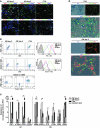
Figure 1. Activated macrophages accumulate and persist in CVUs.
(A) Quantification of CD68+ infiltrating macrophages in NS, AWs at days 1, 2, and 5 after wounding, and CVUs were counted in 10 high-power fields per sample. Results are mean ± SD ratio of CD68+ to total cells counted in the dermis (n = 5). **P < 0.01 versus day-5 AW and NS. (B and C) Representative photomicrographs of skin sections. Activation of macrophages was assessed in NS, AWs, and CVUs by immunostaining of cryosections for the classical activation markers (B) iNOS and (C) TNF-α or the nonclassical activation marker CD163. Nuclei were counterstained with DAPI. Scale bars: 100 μm. Dashed lines indicate the junction between epidermis (e) and dermis. es, eschar; wm, wound margin.
Figure 2
Figure 2. The identified macrophage population mounts an unrestrained proinflammatory M1 activation phenotype and accumulates iron in CVUs.
(A) Representative photomicrographs with double immunostaining of skin cryosections from AWs and CVUs for M1 and M2 macrophage activation markers TNF-α and CD206 or IL-12 and arginase-1. Nuclei were stained with DAPI. Scale bars: 100 μm. Dashed lines indicate the junction between eschar and wound margin. (B) Flow cytometry analysis of wound macrophages purified from AW tissue 2 and 5 days after wounding and CVUs gated according to side scatter (SSC) and CD68 and regated for CD68 and M1 marker TNF-α, CD68 and M2 marker Dectin-1, or TNF-α and Dectin-1. (C) Expression levels for M1 and M2 activation markers of macrophages isolated from 5 AWs and 6 CVUs by flow cytometry. Results are given in RFU (see Methods). Resident skin macrophages were pooled from NS (n ≥ 5). *P < 0.05, **P < 0.01, Student’s t test. (D) Representative photomicrographs of cryosections from CVU patients and AWs from healthy volunteers stained for iron by Perl Prussian blue and immunostained with CD163 for macrophages. Nuclei were stained with PI. High amounts of iron were identified within the CD163+ macrophages (filled arrows) and extracellular space (open arrows) in CVUs, but not AWs. Scale bars: 150 μm.
Figure 3
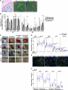
Figure 3. Iron is causal for induction of the macrophage population with unrestrained proinflammatory M1 phenotype and for impaired wound healing.
(A) Representative photomicrographs detects Fe deposition in NS and in F4/80+ macrophages in iron-dextran–treated wounds. Iron deposition was almost completely prevented by coinjection with DFX. Cell nuclei were counterstained with PI. Scale bars: 150 μm. Dashed lines indicate the junction between epidermis and dermis (d). h, hair follicle. (B) Flow cytometry analysis of macrophages isolated from mouse wounds gated for side scatter and F4/80. Expression of M1 and M2 activation markers is shown in RFU (see Methods). Resident skin macrophages were pooled from NS (n = 5). *P < 0.05, **P < 0.01, ***P < 0.001, Student’s t test. (C) Representative macroscopic aspects of wounds at 0, 5, 7, and 10 days after wounding. (D) Statistical analysis of 20 wound areas per group, expressed as percentage of the initial wound size (day 0), for iron-dextran–treated mice (filled symbols) in the presence and absence of DFX or etanercept. Open symbols denote PBS-dextran treatment. Results are mean ± SD (n = 5) representing 1 of 3 independent experiments. *P < 0.05, Mann-Whitney test. (E) Representative photomicrographs of mouse wounds at day 5 after wounding stained for F4/80 and TNF-α, with persistent TNF-α–producing macrophages (yellow), in iron-loaded, but not PBS-dextran– or etanercept-treated, wounds. Nuclei were stained with DAPI. Scale bars: 100 μm. Dashed lines indicate the junction between eschar and wound margin. (F) Statistical analysis of 20 wound areas per group for iron-loaded and PBS-dextran control mice treated with clodronate or PBS liposomes. *P < 0.05, **P < 0.01, Mann-Whitney test.
Figure 4
Figure 4. The iron-induced macrophage population with unrestrained proinflammatory M1 phenotype releases toxic amounts of OH and ONOO in situ.
(A) Representative photomicrographs of skin cryosections derived from wound margins of AWs 2 days after wounding and CVUs. Oxidative burst was detected in cryosections incubated with DHR, a ROS-sensitive dye; thus, ROS concentrations correlated with green fluorescence. Shown are coincubations of cryosections with DHR and SOD, which scavenges O2–•, with the H2O2 scavenger Cat, and with the OH scavenger DMSO. Nuclei were stained with DAPI. Scale bars: 150 μm. (B and C) Representative photomicrographs of paraffin-embedded skin sections from AWs 3 days after wounding and CVUs stained with (B) an antibody against 8OHdG and (C) an antibody against 3-NT. Dashed lines indicate the junction between epidermis and dermis. Scale bars: 150 μm; 50 μm (insets). Quantification of positive cells per 10 high-power fields, assessed for 10 different sections of 5 different AW and CVU samples, is shown as mean ± SD ratios of positive to total cells. (B) The brown-colored precipitate is indicative of oxidative DNA damage in CVUs (inset, arrows), but not AWs. (C) Increased protein nitration was observed in CVUs, but not AWs. (D) Nitroblot analysis of wound lysates with an antibody against 3-NT from PBS-treated mice and from iron-loaded mice and iron-loaded mice treated with DFX, etanercept, or clodronate equilibrated to actin. Positive bands indicate increased levels of protein nitration in iron-loaded wounds compared with reduced levels in iron-dextran–loaded mice treated with DFX, etanercept, or clodronate.
Figure 5
Figure 5. Enhanced ROS release by the unrestrained proinflammatory M1 activated macrophage population activates a senescence program in resident wound fibroblasts.
(A) Representative photomicrographs of cryosections from wound margins from CVUs and NS with increased numbers of γH2AX+ foci in the cell nuclei of wound margins. Double staining for γH2AX and the leukocyte marker CD18 revealed exclusive γH2AX expression in wound-adjacent fibroblasts. Nuclei were stained with DAPI. Scale bars: 150 μm; 50 μm (inset). (B) Quantification of γH2AX+ and γH2AX+CD18+ cells in 10 high-power fields from CVUs and NS samples (n = 5). Results are mean ± SD percent positive cells relative to total cells. **P < 0.01, Student’s t test. (C) Representative Western blot of wound lysates from iron-loaded and PBS control wounds with and without treatment with DFX, equilibrated to total actin, showing expression of γH2AX in iron-loaded, but not control or DFX-treated, wounds (n = 3). (D) Representative photomicrographs of cryosections of wound margins and NS derived from CVU patients showing increased numbers of p16INK4a in the cell nuclei of ulcer margins. Double staining for p16INK4a and CD18 showed p16INK4a expression exclusively in wound-adjacent fibroblasts, not in CD18+ cells. Nuclei were stained with DAPI. Scale bars: 150 μm. Original magnification of insets, ×400. (E) Quantification of p16INK4a+ and p16INK4a+CD18+ cells in 10 high-power fields from 5 different CVUs and NS samples. Results are mean ± SD percent positive cells relative to total cells. **P < 0.01, Student’s t test.
Figure 6
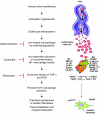
Figure 6. Iron-dependent activation of the macrophage population with unrestrained proinflammatory M1 phenotype, leading to chronic inflammation, tissue breakdown, and impaired wound healing in CVUs.
Chronic venous valve insufficiency leads to hypertension in the lower-limb veins, with persistent erythrocyte extravasation. Engulfment of red blood cells by tissue macrophages (erythrophagocytosis) and release of hemoglobin-bound iron activates a proinflammatory M1 macrophage population with enhanced release of TNF-α, ONOO, and OH. TNF-α expression perpetuates unrestrained proinflammatory M1 activation. ONOO and OH result in oxidative and nitrative damage and induction of a p16INK4a-induced senescent program in wound resident fibroblasts, which therefore cannot contribute to efficient tissue restoration.

Comment in

  • Macrophages: Iron macrophages.
    Papatriantafyllou M. Papatriantafyllou M. Nat Rev Immunol. 2011 Mar;11(3):158. doi: 10.1038/nri2947. Nat Rev Immunol. 2011. PMID: 21452590 No abstract available.

Similar articles

Cited by

References

    1. Bergan JJ, Schmid-Schönbein GW, Smith PD, Nicolaides AN, Boisseau MR, Eklof B. Chronic venous disease. N Engl J Med. 2006;355(5):488–498. doi: 10.1056/NEJMra055289. - DOI - PubMed
    1. Raju S, Neglen P. Clinical practice. Chronic venous insufficiency and varicose veins. N Engl J Med. 2009;360(22):2319–2327. doi: 10.1056/NEJMcp0802444. - DOI - PubMed
    1. Margolis DJ, Bilker W, Santanna J, Baumgarten M. Venous leg ulcer: incidence and prevalence in the elderly. J Am Acad Dermatol. 2002;46(3):381–386. doi: 10.1067/mjd.2002.121739. - DOI - PubMed
    1. Singer AJ, Clark RA. Cutaneous wound healing. . N Engl J Med. 1999;341(10):738–746. doi: 10.1056/NEJM199909023411006. - DOI - PubMed
    1. Agren MS, et al. Causes and effects of the chronic inflammation in venous leg ulcers. Acta Derm Venereol Suppl (Stockh). 2000;210:3–17. - PubMed

Publication types