. 2011 Mar;121(3):956-65.
doi: 10.1172/JCI43475. Epub 2011 Feb 7.

IRBIT governs epithelial secretion in mice by antagonizing the WNK/SPAK kinase pathway

Affiliations

IRBIT governs epithelial secretion in mice by antagonizing the WNK/SPAK kinase pathway

Dongki Yang et al. J Clin Invest. 2011 Mar.

Abstract

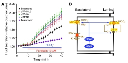
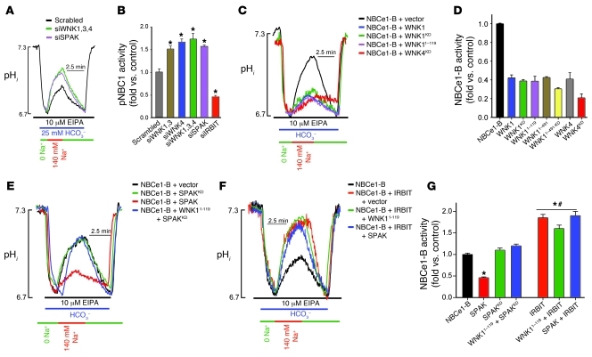
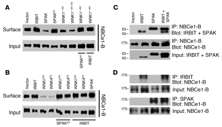
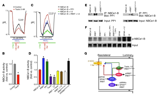
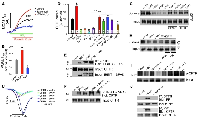
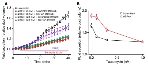
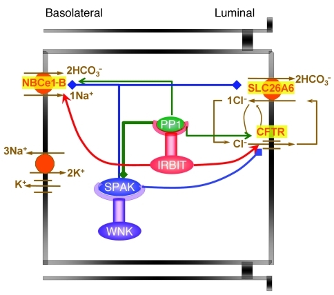
Fluid and HCO(3)(-) secretion are fundamental functions of epithelia and determine bodily fluid volume and ionic composition, among other things. Secretion of ductal fluid and HCO(3)(-) in secretory glands is fueled by Na(+)/HCO(3)(-) cotransport mediated by basolateral solute carrier family 4 member 4 (NBCe1-B) and by Cl(-)/HCO(3)(-) exchange mediated by luminal solute carrier family 26, member 6 (Slc26a6) and CFTR. However, the mechanisms governing ductal secretion are not known. Here, we have shown that pancreatic ductal secretion in mice is suppressed by silencing of the NBCe1-B/CFTR activator inositol-1,4,5-trisphosphate (IP(3)) receptor-binding protein released with IP(3) (IRBIT) and by inhibition of protein phosphatase 1 (PP1). In contrast, silencing the with-no-lysine (WNK) kinases and Ste20-related proline/alanine-rich kinase (SPAK) increased secretion. Molecular analysis revealed that the WNK kinases acted as scaffolds to recruit SPAK, which phosphorylated CFTR and NBCe1-B, reducing their cell surface expression. IRBIT opposed the effects of WNKs and SPAK by recruiting PP1 to the complex to dephosphorylate CFTR and NBCe1-B, restoring their cell surface expression, in addition to stimulating their activities. Silencing of SPAK and IRBIT in the same ducts rescued ductal secretion due to silencing of IRBIT alone. These findings stress the pivotal role of IRBIT in epithelial fluid and HCO(3)(-) secretion and provide a molecular mechanism by which IRBIT coordinates these processes. They also have implications for WNK/SPAK kinase-regulated processes involved in systemic fluid homeostasis, hypertension, and cystic fibrosis.


Figures

Figure 1
Figure 1. The WNK/SPAK and IRBIT/PP1 pathways in ductal fluid secretion.
(A) Sealed pancreatic ducts in primary culture were treated with the indicated siRNA for 48–60 hours before measurement of stimulated ductal fluid secretion by video microscopy. Ducts treated with scrambled siRNA were also treated with the specific PP1 inhibitor tautomycin (3 nM). Similar inhibition of fluid secretion by tautomycin was observed with ducts not treated with scrambled siRNA. Secretion was initiated by incubation of the ducts in HCO3-buffered medium and stimulation with 10 μM forskolin. The traces are the mean ± SEM of 4–10 experiments. (B) Model of the main transporters determining ductal fluid secretion and how they may be regulated by the WNK/SPAK pathway.
Figure 2
Figure 2. The WNK/SPAK pathway inhibits and IRBIT reverses inhibition of NBCe1-B activity.
(A and B) Sealed pancreatic ducts treated with siRNA designed to knock down WNK1+WNK3 (siWNK1,3), WNK4, WNK1+WNK3+WNK4, and IRBIT were used to measure Na+-HCO3 cotransport activity as the Na+-dependent recovery from acid load in the presence of 10 μM EIPA. The results in B are the mean ± SEM of 3–8 ducts obtained from 3 separate mice; *P < 0.01. (CG) HeLa cells were transfected with NBCe1-B and either empty vector (black traces and first bar) or the indicated WNK, SPAK, and IRBIT constructs, and Na+-HCO3 cotransport activity was measured. The bars show the mean ± SEM of 4–8 separate experiments (transfections), and for all conditions in D, P < 0.001 versus control. In G, *P < 0.01 versus control; #P < 0.001 versus the condition without IRBIT.
Figure 3
Figure 3. The WNK/SPAK pathway inhibits and IRBIT reverses inhibition of NBCe1-B surface expression without dissociating the NBCe1-B–SPAK complex.
(A and B) HEK cells were transfected with GFP–NBCe1-B and the indicated WNK and Flag-SPAK constructs. Note that in the last two lanes in A, WNK11–491 was coexpressed with SPAKKD and with IRBIT; and in lanes 5, 6 and 7, 8 in B, WNK1KD and WNK4KD were coexpressed with Flag-SPAKKD and Flag-IRBIT, respectively. In lane 9 Flag-SPAK was coexpressed with Flag-IRBIT. The cells were used to determine total (lower blots) and surface expression (upper blots) of GFP–NBCe1-B by biotinylation. (C and D) HEK cells were transfected with GFP–NBCe1-B and with empty vector, Flag-IRBIT, Flag-SPAK, or Flag-IRBIT+Flag-SPAK and were used to determine the effect of IRBIT and SPAK on their interaction with GFP–NBCe1-B by coimmunoprecipitation assays. Note that IRBIT and SPAK do not affect their mutual interaction with NBCe1-B. NBCe1-B was detected with anti-GFP and SPAK and IRBIT with anti-Flag antibodies. Analysis of multiple experiments and averages are given in Supplemental Figure 3.
Figure 4
Figure 4. PP1 mediates all effects of IRBIT.
(A and B) Sealed pancreatic ducts were treated with vehicle or with 3 nM tautomycin (Tauto) for 10 minutes, and Na+-HCO3 cotransport activity was measured. B shows mean ± SEM (n = 3, *P < 0.001). (C and D) HeLa cells transfected with NBCe1-B and vector (black), PP1 (red), inhibitor-2 (I-2; green), or inhibitor-2 plus IRBIT (blue). Cells expressing NBCe1-B were also transfected with IRBIT (bars 2 and 6), IRBITI42F44/AA (bar 8), and 3XIRBITI42F44/AA (bar 9) and treated with 3 nM tautomycin for 10 minutes (bars 4 and 6). D shows mean ± SEM of Na+-HCO3 cotransport (n = 4–8, *P < 0.01). (E) HEK cells transfected with NBCe1-B and vector, IRBIT, or IRBITI42F44/AA were used to test recruitment of the native PP1 to NBCe1-B. (F) HEK cells transfected with NBCe1-B and the indicated combinations of IRBIT, PP1, inhibitor-2, and SPAK constructs were labeled with 32P for 2 hours. NBCe1-B was immunoprecipitated and NBCe1-B phosphorylation tested by radiography. (G) Model of regulation of NBCe1-B surface expression and activity by the WNK/SPAK and IRBIT/PP1 pathways and the relationship between them. The WNKs function as scaffolds for SPAK, which phosphorylates NBCe1-B to reduce its surface expression, and IRBIT functions as a scaffold for PP1, which dephosphorylates NBCe1-B to restore surface expression. IRBIT also activates NBCe1-B by preventing autoinhibition by NBCe1-B N-terminus. The black lines represent the probes used to test each component of the pathways. Averages for E and F are given in Supplemental Figure 4A and Supplemental Figure 5A, respectively.
Figure 5
Figure 5. Regulation of CFTR by the WNK/SPAK and IRBIT/PP1 pathways.
(A and B) Sealed ducts were treated with scrambled siRNA (black, blue) or WNK (red) siRNA and incubated for 5 minutes with 3 nM tautomycin (blue). CFTR activity was measured with MQAE as NO3/Cl exchange activity. B shows mean ± SEM (n = 3, *P < 0.01). (C and D) CFTR current was measured in HEK cells transfected with CFTR and the indicated IRBIT, WNK, and SPAK combinations or treated with 3 nM tautomycin for 5 minutes. D shows mean ± SEM (n = 4–17; *P < 0.01; #P < 0.05 versus control). Currents were normalized to cell capacitance before averaging. (E and F) Mutual interaction of IRBIT and SPAK with CFTR, as measured by the reciprocal coimmunoprecipitation assays. In F, asterisks in lane 4 indicate expression of 4 times excess SPAK relative to IRBIT and in lane 5 expression of 4 times excess IRBIT relative to SPAK. (G) HEK cells expressing CFTR and the indicated combinations of IRBIT, WNK1, WNK4, and SPAK constructs were used to test their effect on surface expression of CFTR by biotinylation. (H) Inhibition of CFTR surface expression by WNK4, WNK1, and WNK11–119 and its reversal by SPAKKD and IRBIT. (I) HEK cells expressing CFTR and the indicated combinations of IRBIT, IRBITI42F44/AA, PP1, SPAK, and inhibitor-2 were used to measure CFTR phosphorylation, as described in Figure 3 for NBCe1-B. (J) HEK cells expressing CFTR and cotransfected with IRBIT or IRBITI42F44/AA were used to show IRBIT-mediated recruitment of the native PP1 to CFTR. Averages for G and H are given in Supplemental Figure 4B and for I in Supplemental Figure 5B.
Figure 6
Figure 6. Physiological relevance of regulation of the WNK/SPAK pathway by the IRBIT/PP1 pathway.
(A) Sealed pancreatic ducts were treated with 10 nM scrambled siRNA (black, red, and green traces), 5 nM IRBIT siRNA (red and blue traces), 2.5 nM IRBIT siRNA (green and purple traces), and 10 nM SPAK siRNA (blue and purple traces) and were used to measure stimulated ductal fluid secretion. (B) Sealed pancreatic ducts were treated with 10 nM scrambled (black trace) or SPAK siRNA (red trace). Ten minutes before and during the experiments, the ducts were treated with the indicated concentration of tautomycin, and fluid secretion was measured by video microscopy for 40 minutes. Results in A and B are mean ± SEM of 3–6 experiments.
Figure 7
Figure 7. Regulation of ductal secretion by the WNK/SPAK and the IRBIT/PP1 pathways.
The model shows that the WNK/SPAK pathway determines the duct resting secretory state by reducing NBCe1-B and CFTR surface expression and thus their activity. The WNKs are modeled to function as scaffolds for SPAK, and SPAK phosphorylates NBCe1-B and CFTR (blue lines). IRBIT determines the secretory state and dominates ductal secretion by both reversing the effect of the WNK/SPAK pathway and directly activating NBCe1-B and CFTR. IRBIT functions as a scaffold for PP1 to recruit PP1 to a complex with NBCe1-B and CFTR to dephosphorylate NBCe1-B and CFTR and restore their surface expression (green arrows). IRBIT then directly activates NBCe1-B and CFTR by preventing NBCe1-B autoinhibition by its N-terminal domain (24) and by interaction of the IRBIT PEST domain with CFTR (21). The dual effects of IRBIT, reversing the inhibitory state and stimulating the transporters, highlight the prominent role of IRBIT in epithelial fluid and electrolyte secretion.

Similar articles

Cited by

References

    1. Melvin JE, Yule D, Shuttleworth T, Begenisich T. Regulation of fluid and electrolyte secretion in salivary gland acinar cells. Annu Rev Physiol. 2005;67:445–469. doi: 10.1146/annurev.physiol.67.041703.084745. - DOI - PubMed
    1. Steward MC, Ishiguro H, Case RM. Mechanisms of bicarbonate secretion in the pancreatic duct. Annu Rev Physiol. 2005;67:377–409. doi: 10.1146/annurev.physiol.67.031103.153247. - DOI - PubMed
    1. Kahle KT, Ring AM, Lifton RP. Molecular physiology of the WNK kinases. Annu Rev Physiol. 2008;70:329–355. - PubMed
    1. Dorwart MR, Shcheynikov N, Yang D, Muallem S. The solute carrier 26 family of proteins in epithelial ion transport. Physiology (Bethesda). 2008;23:104–114. - PubMed
    1. Seidler U, et al. Knockout mouse models for intestinal electrolyte transporters and regulatory PDZ adaptors: new insights into cystic fibrosis, secretory diarrhoea and fructose-induced hypertension. Exp Physiol. 2009;94(2):175–179. doi: 10.1113/expphysiol.2008.043018. - DOI - PubMed

Publication types

MeSH terms