Identification and characterization of three novel small interference RNAs that effectively down-regulate the isolated nucleocapsid gene expression of SARS coronavirus
- PMID: 21317844
- PMCID: PMC6259856
- DOI: 10.3390/molecules16021544
Identification and characterization of three novel small interference RNAs that effectively down-regulate the isolated nucleocapsid gene expression of SARS coronavirus
Abstract
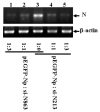
Nucleocapsid (N) protein of severe acute respiratory syndrome-associated coronavirus (SARS-CoV) is a major pathological determinant in the host that may cause host cell apoptosis, upregulate the proinflammatory cytokine production, and block innate immune responses. Therefore, N gene has long been thought an ideal target for the design of small interference RNA (siRNA). siRNA is a class of small non-coding RNAs with a size of 21-25nt that functions post-transcriptionally to block targeted gene expression. In this study, we analyzed the N gene coding sequences derived from 16 different isolates, and found that nucleotide deletions and substitutions are mainly located at the first 440nt sequence. Combining previous reports and the above sequence information, we create three novel siRNAs that specifically target the conserved and unexploited regions in the N gene. We show that these siRNAs could effectively and specifically block the isolated N gene expression in mammal cells. Furthermore, we provide evidence to show that N gene can effectively up-regulate M gene mediated interferon b (IFNb) production, while blocking N gene expression by specific siRNA significantly reduces IFNb gene expression. Our data indicate that the inhibitory effect of siRNA on the isolated N gene expression might be influenced by the sequence context around the targeted sites.
Figures


Similar articles
-
Small interfering RNA effectively inhibits the expression of SARS coronavirus membrane gene at two novel targeting sites.Molecules. 2010 Oct 18;15(10):7197-207. doi: 10.3390/molecules15107197. Molecules. 2010. PMID: 20956884 Free PMC article.
-
Small interfering RNA inhibits SARS-CoV nucleocapsid gene expression in cultured cells and mouse muscles.FEBS Lett. 2005 Apr 25;579(11):2404-10. doi: 10.1016/j.febslet.2005.02.080. FEBS Lett. 2005. PMID: 15848179 Free PMC article.
-
Inhibition of genes expression of SARS coronavirus by synthetic small interfering RNAs.Cell Res. 2005 Mar;15(3):193-200. doi: 10.1038/sj.cr.7290286. Cell Res. 2005. PMID: 15780182 Free PMC article.
-
siRNA targeting the leader sequence of SARS-CoV inhibits virus replication.Gene Ther. 2005 May;12(9):751-61. doi: 10.1038/sj.gt.3302479. Gene Ther. 2005. PMID: 15772689 Free PMC article.
-
Assembly of severe acute respiratory syndrome coronavirus RNA packaging signal into virus-like particles is nucleocapsid dependent.J Virol. 2005 Nov;79(22):13848-55. doi: 10.1128/JVI.79.22.13848-13855.2005. J Virol. 2005. PMID: 16254320 Free PMC article.
Cited by
-
Non-Coding RNAs and SARS-Related Coronaviruses.Viruses. 2020 Dec 1;12(12):1374. doi: 10.3390/v12121374. Viruses. 2020. PMID: 33271762 Free PMC article. Review.
-
Development and application of ribonucleic acid therapy strategies against COVID-19.Int J Biol Sci. 2022 Aug 1;18(13):5070-5085. doi: 10.7150/ijbs.72706. eCollection 2022. Int J Biol Sci. 2022. PMID: 35982905 Free PMC article. Review.
-
Prospects for RNAi Therapy of COVID-19.Front Bioeng Biotechnol. 2020 Jul 30;8:916. doi: 10.3389/fbioe.2020.00916. eCollection 2020. Front Bioeng Biotechnol. 2020. PMID: 32850752 Free PMC article. Review.
References
-
- Ruan Y.J., Wei C.L., Ee A.L., Vega V.B., Thoreau H., Su S.T., Chia J.M., Ng P., Chiu K.P., Lim L., et al. Comparative full-length genome sequence analysis of 14 SARS coronavirus isolates and common mutations associated with putative origins of infection. Lancet. 2003;361:1779–1785. doi: 10.1016/S0140-6736(03)13414-9. - DOI - PMC - PubMed
-
- Hatakeyama S., Matsuoka Y., Ueshiba H., Komatsu N., Itoh K., Shichijo S., Kanai T., Fukushi M., Ishida I., Kirikae T., et al. Dissection and identification of regions required to form pseudoparticles by the interaction between the nucleocapsid (N) and membrane (M) proteins of SARS coronavirus. Virology. 2008;380:99–108. doi: 10.1016/j.virol.2008.07.012. - DOI - PMC - PubMed
Publication types
MeSH terms
Substances
LinkOut - more resources
Full Text Sources
Miscellaneous

