. 2011 Mar 16;30(6):1079-92.
doi: 10.1038/emboj.2011.27. Epub 2011 Feb 11.

Factors determining DNA double-strand break repair pathway choice in G2 phase

Affiliations

Factors determining DNA double-strand break repair pathway choice in G2 phase

Atsushi Shibata et al. EMBO J. .

Abstract

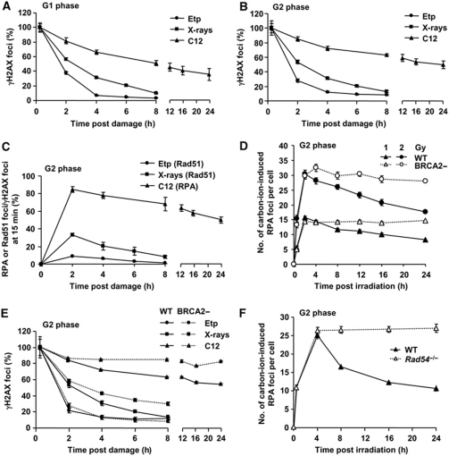
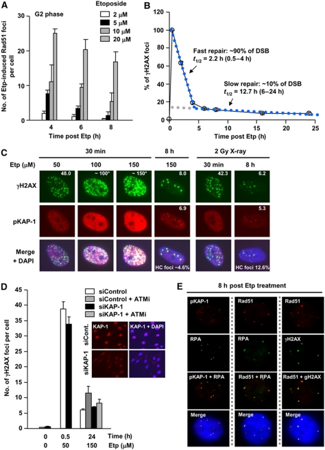
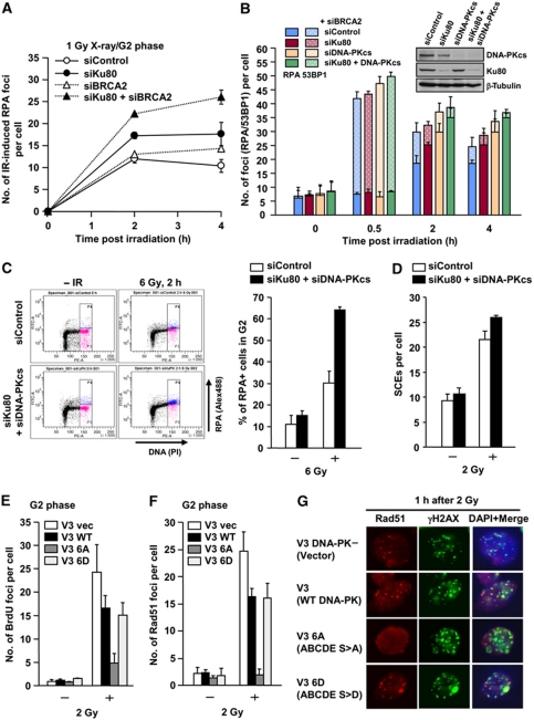
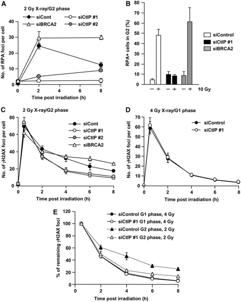
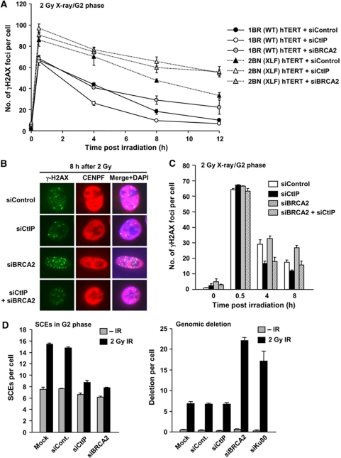
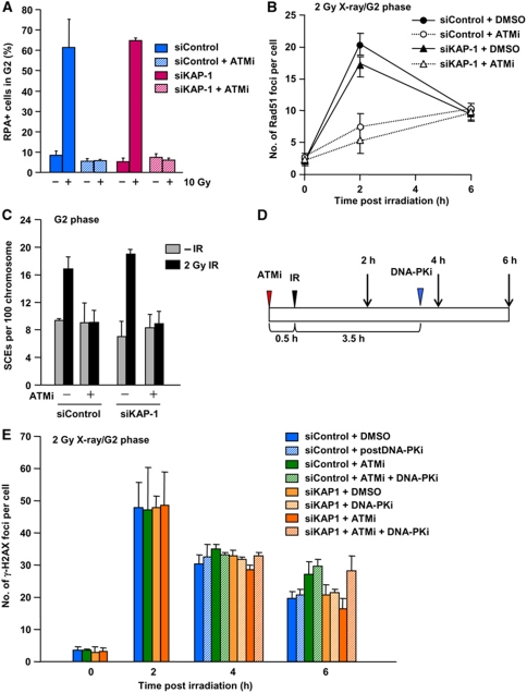
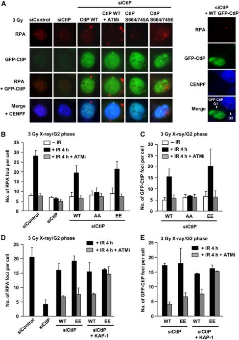
DNA non-homologous end joining (NHEJ) and homologous recombination (HR) function to repair DNA double-strand breaks (DSBs) in G2 phase with HR preferentially repairing heterochromatin-associated DSBs (HC-DSBs). Here, we examine the regulation of repair pathway usage at two-ended DSBs in G2. We identify the speed of DSB repair as a major component influencing repair pathway usage showing that DNA damage and chromatin complexity are factors influencing DSB repair rate and pathway choice. Loss of NHEJ proteins also slows DSB repair allowing increased resection. However, expression of an autophosphorylation-defective DNA-PKcs mutant, which binds DSBs but precludes the completion of NHEJ, dramatically reduces DSB end resection at all DSBs. In contrast, loss of HR does not impair repair by NHEJ although CtIP-dependent end resection precludes NHEJ usage. We propose that NHEJ initially attempts to repair DSBs and, if rapid rejoining does not ensue, then resection occurs promoting repair by HR. Finally, we identify novel roles for ATM in regulating DSB end resection; an indirect role in promoting KAP-1-dependent chromatin relaxation and a direct role in phosphorylating and activating CtIP.


Conflict of interest statement

The authors declare that they have no conflict of interest.

Figures

Figure 1
Figure 1
DSB end complexity determines the speed of DSB repair and the level of DSB end resection in G2 phase. (A, B) Positive correlation between DNA damage complexity and repair kinetics. The kinetics of DSB repair following exposure to 2 Gy carbon ions, 2 Gy X-rays and 20 (A) or 5 (B) μM Etp for 15 min, was monitored by enumerating γH2AX foci in HSF1 primary cells in G1 (A) or G2 (B) cells. The percent of γH2AX foci is normalised to γH2AX foci numbers at 15 min after damage (representing the number of DSBs induced). The dose of Etp chosen produced a similar number of DSBs to that induced by 2 Gy X-rays. Irradiated G1 and G2 cells were identified as CENP-F negative and positive, respectively. S phase cells identified by pan-nuclear γH2AX staining were excluded from analysis (Supplementary Figure S1). (C) The magnitude of resection correlates with the speed of DSB repair. RPA or Rad51 foci numbers were enumerated following exposure to 2 Gy carbon ions, 2 Gy X-rays and 5 μM Etp in HSF1 primary cells. The number of RPA or Rad51 foci at the times indicated was normalised to γH2AX foci numbers at 15 min after damage. RPA and Rad51 foci numbers were similar (data not shown). The actual numbers of γH2AX and RPA–Rad51 foci scored per cell in G2 phase are given in Supplementary Figure S3C and D. We have previously undertaken kinetic analyses of RPA foci formation and shown maximal numbers 2 h post-IR (Beucher et al, 2009). Here, we observe a similar finding for Rad51 foci formation (Supplementary Figure S2C). Further discussion of this analysis is given in Supplementary Figure S2 legend. (D) Total DSB end resection events in HSF1 (WT) and HSC62 (BRCA2) cells. Following exposure to 1 or 2 Gy carbon ions, the number of RPA foci was scored in G2 cells. (E) The contribution of BRCA2-dependent HR to G2 DSB repair following exposure to 2 Gy carbon ions, 2 Gy X-rays and 5 μM Etp. Approximately 90% of DSBs remained in HSC62 (BRCA2) cells at 24 h after carbon ions compared with ∼30% of X-ray-induced DSBs. There is no detectable DSB repair defect in BRCA2-deficient cells following exposure to Etp. (F) More than ∼90% of RPA foci persist in Rad54−/− MEFs up to 24 h after carbon-ion irradiation. The number of RPA foci was enumerated after 2 Gy carbon ions. G2 cells were identified as speckled p-histoneH3 Ser10 staining. Error bars represent the s.e.m. of three experiments.
Figure 2
Figure 2
Slowly repaired etoposide-induced DSBs localise to heterochromatin and undergo resection. (A) Rad51 foci arise following exposure to Etp for 30 min in G2 cells in a dose-dependent manner. Cells were exposed to increasing doses of Etp and Rad51 foci enumerated in HSF1 primary cells. The number of Rad51 foci in untreated cells (<2) was subtracted from results shown. (B) DSB repair kinetics in 48BR primary G0/G1 cells following exposure to 50 μM Etp for 30 min. Approximately 90% of DSBs are repaired with fast kinetics from 0.5 to 4 h (half life: 2.2 h); ∼10% of DSBs are repaired with slower kinetics from 6 to 24 h (half life: 12.7 h). Direct comparison of repair kinetics between Etp and IR is shown in Supplementary Table S1. (C) A low fraction of HC-associated DSBs arise following Etp treatment. 48BR primary G0/G1 cells were treated with 50, 100 and 150 μM Etp and foci enumerated at 30 min and 8 h. Total DSBs were detected with γH2AX foci, whereas HC-associated DSBs were detected with pKAP-1 foci. (N.B. after DSB induction at 30 min, pan-nuclear KAP-1 phosphorylation obscures the presence of pKAP-1 foci; thus foci were only scored at 8 h (Noon et al, 2010)). DSB induction was estimated from γH2AX present at 30 min assuming a dose-linear induction rate. The HC-associated DSBs at 8 h after Etp treatment was approximately three-fold lower than following X-ray exposure, 4.6 and 12.6%, respectively. (D) ATM-dependent DSB repair after Etp treatment is alleviated by KAP-1 siRNA. 48BR primary cells were exposed to 50 and 150 μM Etp for 30 min with or without ATMi. ATMi was readded after medium refreshing. γH2AX foci enumerated at 30 min and 24 h in G1 phase. Right panel shows KAP-1 knockdown efficiency. (E) RPA, Rad51 and γH2AX foci in Etp-treated G2 cells co-localise with the HC-DSB marker, pKAP-1. 48BR primary cells were exposed to 10 μM Etp and fixed 8 h after treatment. In these experiments (AE), Etp was added for 30 min in medium at 37°C. Cells were washed with PBS three times before adding drug-free medium and cells were then incubated until the indicated time points.
Figure 3
Figure 3
DNA-PK competes with DSB end resection. (A) Ku80 siRNA significantly increases RPA foci numbers post-IR. A549 cells were exposed to 1 Gy X-rays and RPA foci were enumerated as indicated either with or without BRCA2 siRNA. G2 cells were identified with CENP-F. Knockdown efficiency and typical images are shown in Supplementary Figure S5A. (B) Ku80 siRNA, DNA-PKcs siRNA or combined siRNA causes increased DSB end resection in A549 G2 cells. RPA and 53BP1 foci were enumerated after 1 Gy X-rays. Solid bars represent the number of RPA foci, and solid+hashed bars represent the total number of 53BP1 foci. The knockdown efficiencies are shown in the right panel. (C) Loss of Ku80 and DNA-PKcs increased IR-induced RPA retention in G2. RPA retention in G2 cells was analysed using α-RPA antibody after detergent extraction. Chromatin-associated RPA was detected as an Alex488 signal following FACS. APH alone does not induce detectable RPA signal by FACS (data not shown), although some level of RPA signal was detected by IF (Supplementary Figure S2). Percent of RPA positive was shown in the right panel. Error bars represent two independent experiments. (D) Increased IR-induced SCEs in Ku80 and DNA-PKcs double-knockdown G2 cells. IR-induced SCEs in G2 phase were analysed following 2 Gy X-rays. (E) A significant reduction of IR-induced BrdU foci formation in CHO cells expressing DNA-PKcs ABCDE S>A. In all, 20 μM BrdU was added 24 h before IR. Cells were extracted with 0.2% Triton for 1 min at 2 h after 2 Gy and stained with α-BrdU antibody without denaturation. (F, G) CHO cells expressing DNA-PKcs ABCDE S>A fail to form Rad51 foci at 1 h after 2 Gy X-rays in contrast to control cells and cells expressing the phosphorylation mimic, DNA-PKcs ABCDE S>D. G2 cells were identified by DAPI intensity using ImageJ. Similar results were obtained examining RPA foci (data not shown). DNA-PK expression levels in the V3 strains are shown in Supplementary Figure S5B. Further characterisation of the strains has been described previously (Chan and Lees-Miller, 1996; Cui et al, 2005; Meek et al, 2007).
Figure 4
Figure 4
CtIP siRNA-treated G2 cells show faster DSB repair kinetics compared with control cells but similar to G1 phase DSB repair. (A) CtIP siRNA treatment abolishes DSB end resection, whereas RPA foci levels are sustained following BRCA2 siRNA. A549 cells were treated with control, BRCA2 or CtIP siRNA. RPA foci were enumerated after 2 Gy X-rays. Similar results were obtained using two distinct CtIP siRNA oligonucleotides. (B) Cells subjected to CtIP siRNA show impaired IR-induced RPA retention. RPA retention was analysed as described in Figure 3C. Consistent with RPA foci data, CtIP siRNA cells show loss of RPA signal. (C) CtIP siRNA cells show faster repair kinetics at 4–8 h compared with control cells, whereas BRCA2 siRNA cells show a DSB repair defect. Following exposure to 2 Gy X-rays, DSB repair kinetics in A549 G2 cells was measured by enumerating γH2AX foci. G2 cells were identified using CENP-F. (D) The kinetics of γH2AX disappearance is unaffected by CtIP siRNA after 4 Gy X-rays. A549 G1 cells were identified as CENP-F negative. (E) Direct comparison of DSB repair kinetics in A549 G1 and G2 cells following control and CtIP siRNA. CtIP siRNA in G2 results in similar repair kinetics to G1 phase, suggesting that DSB repair in G2 following CtIP siRNA occurs by NHEJ. Note that even though all DSBs are repaired by NHEJ, fast and slow kinetics are observed since HC-DSBs are repaired slowly even in G1. In all panels, error bars represent the mean and s.d. from three independent experiments except panel (B), which is from two experiments.
Figure 5
Figure 5
CtIP siRNA allows repair to be switched from HR to NHEJ. (A) CtIP siRNA and BRCA2 siRNA causes a similar DSB repair defect in XLF-defective cells following IR. DSB repair was monitored by γH2AX foci enumeration after 2 Gy X-rays using the treatments and/or cells indicated. As in Figure 4, CtIP siRNA shows faster repair, whereas BRCA2 siRNA confers a repair defect in WT G2 cells. In contrast, CtIP or BRCA2 siRNA in XLF cells showed a similar DSB repair defect, demonstrating that DSB repair following CtIP siRNA occurs by NHEJ. (B, C) CtIP siRNA alleviates the BRCA2-dependent repair defect in A549 G2 cells after 2 Gy X-rays. The slightly higher γH2AX foci numbers following combined BRCA2 and CtIP siRNA than following CtIP siRNA alone at 8 h is probably a result of imperfect siRNA efficiency. (D) CtIP or BRCA2 siRNA inhibits IR-induced SCEs. Ku80 or BRCA2 siRNA but not CtIP siRNA causes increased deletion events. IR-induced SCEs in G2 phase were analysed following 2 Gy X-rays (left panel). Genomic deletions were examined in metaphase preparations following 2 Gy X-rays (right panel).
Figure 6
Figure 6
KAP-1 siRNA does not rescue DSB end resection nor IR-induced SCEs in the presence of ATMi, but DSB repair occurs by NHEJ. (A) ATMi treatment abolishes RPA retention after IR. Additionally, KAP-1 siRNA does not restore RPA retention in the presence of ATMi. Error bars represent the s.d. from two independent experiments. (B) ATMi treatment significantly reduced Rad51 foci numbers after X-ray exposure irrespective of KAP-1 status. Similar results were obtained for RPA foci (data not shown). (C) ATMi treatment abolishes IR-induced SCEs in G2 irrespective of KAP-1 status. IR-induced SCEs in G2 phase were analysed following KAP-1 siRNA with/without ATMi after 2 Gy X-rays. KAP-1 siRNA does not restore Rad51 or SCE formation in the presence of ATMi. (D) Diagram showing the procedure used to examine the role of NHEJ in DSB repair. A549 cells were treated with a DNA-PK inhibitor (DNA-PKi) at 3.5 h post-IR, when most NHEJ-dependent DSB repair is completed after 2 Gy X-rays. (E) IR-induced DSBs are repaired by NHEJ following KAP-1 siRNA in the presence of ATMi. A549 cells were exposed to 2 Gy X-rays. ATMi-treated cells showed a repair defect. Addition of DNA-PKi at 3.5 h post-IR does not affect DSB repair in a control background. KAP-1 siRNA alleviates ATM-dependent DSB repair in G2 (i.e. enhanced repair is observed following KAP-1 siRNA+ATMi) (despite the lack of Rad51 foci formation and SCEs). DNA-PKi addition at 3.5 h prevents the enhanced repair observed following KAP-1 siRNA+ATMi between 4 and 6 h, indicating that the DSBs are repaired by NHEJ.
Figure 7
Figure 7
ATM-dependent CtIP phosphorylation is required for DSB end resection in G2 cells. (A) Typical images of RPA and GFP-CtIP foci at 4 h in U2OS G2 cells after 3 Gy X-rays. In contrast, GFP-CtIP expressed in G1 cells does not form either RPA or GFP-CtIP foci (right panel). CtIP mutant proteins were expressed following CtIP siRNA. (B, C) CtIP siRNA abolished IR-induced RPA foci formation. GFP-CtIP was overexpressed in U2OS cells. GFP-CtIP S664/745A expressing cells fail to form RPA and GFP-CtIP foci after 3 Gy X-rays, whereas wild type and S664/745E GFP-CtIP overexpression rescued RPA/GFP-CtIP foci formation. (D, E) KAP-1 siRNA alleviates loss of GFP-CtIP and RPA foci formation in S664/745E mutant in the presence of ATMi following exposure to 3 Gy X-rays. GFP-CtIP was overexpressed in U2OS cells. KAP-1 siRNA does not affect GFP-CtIP or RPA foci formation in CtIP WT expressing cells, whereas it relieves DSB end resection in S664/745E expressing cells.

Similar articles

Cited by

References

    1. Aylon Y, Liefshitz B, Kupiec M (2004) The CDK regulates repair of double-strand breaks by homologous recombination during the cell cycle. EMBO J 23: 4868–4875 - PMC - PubMed
    1. Beucher A, Birraux J, Tchouandong L, Barton O, Shibata A, Conrad S, Goodarzi AA, Krempler A, Jeggo PA, Lobrich M (2009) ATM and Artemis promote homologous recombination of radiation-induced DNA double-strand breaks in G2. EMBO J 28: 3413–3427 - PMC - PubMed
    1. Chan DW, Lees-Miller SP (1996) The DNA-dependent protein kinase is inactivated by autophosphorylation of the catalytic subunit. J Biol Chem 271: 8936–8941 - PubMed
    1. Cui X, Yu Y, Gupta S, Cho YM, Lees-Miller SP, Meek K (2005) Autophosphorylation of DNA-dependent protein kinase regulates DNA end processing and may also alter double-strand break repair pathway choice. Mol Cell Biol 25: 10842–10852 - PMC - PubMed
    1. Drouet J, Delteil C, Lefrancois J, Concannon P, Salles B, Calsou P (2005) DNA-dependent protein kinase and XRCC4-DNA ligase IV mobilization in the cell in response to DNA double strand breaks. J Biol Chem 280: 7060–7069 - PubMed

Publication types

Substances