. 2011 Mar 16;30(6):1027-39.
doi: 10.1038/emboj.2011.32. Epub 2011 Feb 11.

Red1 promotes the elimination of meiosis-specific mRNAs in vegetatively growing fission yeast

Affiliations

Red1 promotes the elimination of meiosis-specific mRNAs in vegetatively growing fission yeast

Tomoyasu Sugiyama et al. EMBO J. .

Abstract

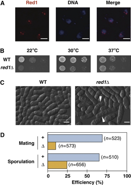
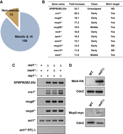
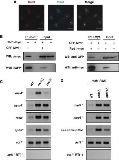
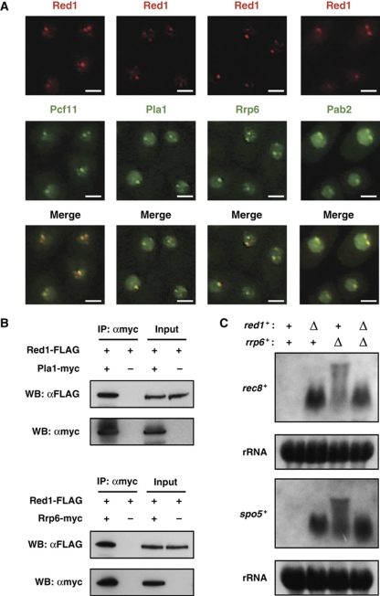
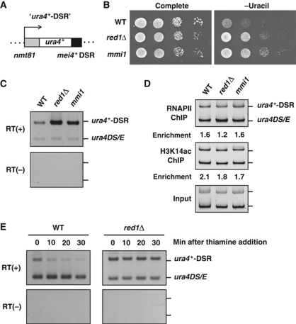
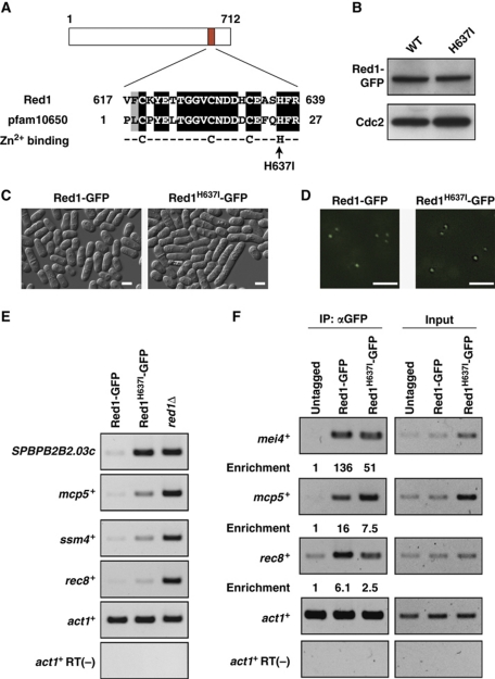
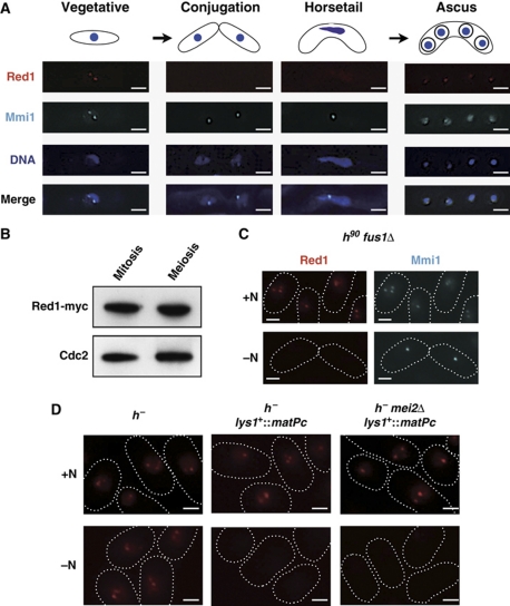
Meiosis-specific mRNAs are transcribed in vegetative fission yeast, and these meiotic mRNAs are selectively removed from mitotic cells to suppress meiosis. This RNA elimination system requires degradation signal sequences called determinant of selective removal (DSR), an RNA-binding protein Mmi1, polyadenylation factors, and the nuclear exosome. However, the detailed mechanism by which meiotic mRNAs are selectively degraded in mitosis but not meiosis is not understood fully. Here we report that Red1, a novel protein, is essential for elimination of meiotic mRNAs from mitotic cells. A red1 deletion results in the accumulation of a large number of meiotic mRNAs in mitotic cells. Red1 interacts with Mmi1, Pla1, the canonical poly(A) polymerase, and Rrp6, a subunit of the nuclear exosome, and promotes the destabilization of DSR-containing mRNAs. Moreover, Red1 forms nuclear bodies in mitotic cells, and these foci are disassembled during meiosis. These results demonstrate that Red1 is involved in DSR-directed RNA decay to prevent ectopic expression of meiotic mRNAs in vegetative cells.


Conflict of interest statement

The authors declare that they have no conflict of interest.

Figures

Figure 1
Figure 1
Red1 localizes as nuclear dots and is required for normal growth and meiotic processes. (A) Red1 forms nuclear bodies in vegetatively growing cells. Yeast cells expressing Red1 tagged with tdTomato were fixed and stained with DAPI. Representative deconvolved images are shown. Bars, 2 μm. (B) red1Δ cells display growth retardation. Serial dilutions of wild-type and red1Δ cells were spotted onto YEA plates and then incubated at various temperatures for 3–5 days before taking pictures. (C) Some red1Δ cells display abnormal cell morphology. Differential interference contrast (DIC) images of wild type (WT) and red1Δ show that red1Δ cells are elongated relative to wild-type cells. Arrow heads indicate cells with abnormalities, including multi-septa and altered cell polarity. Bars, 5 μm. (D) Mating and sporulation efficiencies are reduced in red1Δ cultures. The efficiencies of mating and sporulation in wild-type and red1Δ cultures were measured as described in the Materials and methods.
Figure 2
Figure 2
Meiosis-specific mRNAs are downregulated by Red1 in mitotic cells. (A) A large fraction of the mRNAs accumulating in growing red1Δ are meiotic mRNAs. Expression analyses using a microarray technique demonstrate that 88% of increased (more than two-fold) transcripts have previously been reported as genes upregulated in response to nitrogen starvation/pheromone and during meiosis. (B) The 11 mRNAs exhibiting the greatest increase in red1Δ cells. These genes were classified according to the timing of gene expression in meiosis (Mata et al, 2002). The fold increases shown in the list are the averages of two independent experiments. The Mmi1 target genes have been identified previously (Harigaya et al, 2006). (C) Meiotic mRNAs accumulated in red1Δ cells independently of both Mei4 and Rep1 transcription factors. Total RNA samples from wild-type, red1Δ, red1Δmei4Δ, and red1Δrep1Δ cells were subjected to RT–PCR analyses. RT–PCR of SPBPB2B2.03c, crs1+, mug9+, and mcp5+ indicates that the increased levels of these meiotic transcripts also accumulate in red1Δmei4Δ and red1Δrep1Δ cells. (D) Mei4 and Mcp5 proteins are produced in mitotically growing red1Δ cells. Protein extracts of mitotic wild-type and red1Δ cells expressing Mei4-HA and Mcp5-myc were resolved on a polyacrylamide gel and then analysed by western blotting using anti-HA (top panels) and anti-myc (bottom panels) antibodies. Cdc2 was monitored as a loading control.
Figure 3
Figure 3
Red1 co-localizes and interacts with Mmi1 in vegetative cells. (A) Red1 foci coincide with Mmi1 dots. Deconvolved images of a strain expressing both Red1-tdTomato and CFP-Mmi1 are shown. Bars, 2 μm. (B) Red1 co-immunoprecipitates with Mmi1. Total cell lysates from strains expressing CFP-Mmi1, Red1-myc, and Red1-myc/CFP-Mmi1 were subjected to immunoprecipitation, followed by western blotting. (C) Red1 and Mmi1 have common target genes. RT–PCR of four DSR-containing genes (mei4+, ssm4+, rec8+, and spo5+) indicates that either a red1 deficiency or a mmi1 mutation (mmi1-619) resulted in increased levels of these meiotic mRNAs. (D) Meiotic mRNAs accumulate in conditional mmi1Δ cells. Total RNA samples from mei4-P527, red1Δmei4-P527, and mmi1Δmei4-P527 cells were subjected to RT–PCR analyses. RT–PCR of mei4+, mcp5+, and SPBPB2B2.03c indicates that red1Δ or mmi1Δ in a mei4-P527 background results in increased levels of these meiotic transcripts. Note that mmi1+ was deleted in the mei4-P527 mutant strain because mei4 deficiency suppresses the severe growth defect caused by mmi1Δ.
Figure 4
Figure 4
Red1 localizes to cleavage bodies in mitotically dividing cells and cooperates with polyadenylation factors to hyperadenylate meiotic mRNAs. (A) Red1 dots co-localize with cleavage factor, Pcf11; the canonical poly(A) polymerase, Pla1; a nuclear exosome subunit, Rrp6; and a nuclear poly(A)-binding protein, Pab2. Microscopic images of strains expressing both Red1-tdTomato and either Pcf11-, Pla1-, Rrp6-, or Pab2-GFP are shown. Bars, 2 μm. (B) Red1 co-immunoprecipitates with Pla1 and Rrp6. Total cell extracts prepared from strains expressing both Red1-FLAG and Rrp6/Pla1-myc were subjected to immunoprecipitation, followed by western blotting. (C) Meiotic mRNAs in red1Δ do not have highly elongated poly(A) tails. Northern blotting was performed to analyse rec8+ and spo5+ mRNAs in wild type (WT), red1Δ, rrp6Δ, and red1Δrrp6Δ. The same blots were stained with methylene blue to visualize ribosomal RNAs (rRNA), which serve as a loading control.
Figure 5
Figure 5
Red1 promotes the destabilization of DSR-containing mRNA. (A) Schematic representation of the ura4+-DSR construct. The DSR region derived from mei4+ was fused to the ura4+ gene and this ura4+-DSR mRNA was driven by the nmt81 promoter, which was repressed in the presence of thiamine. This construct was integrated into the lys1+ locus. (B) red1Δ led to the ‘ura4+-on’ state in cells carrying the ura4+-DSR construct. Serial dilutions of wild-type (WT), red1Δ, and mmi1-619 (mmi1) cells containing the ura4+-DSR construct were spotted onto a complete or uracil-lacking plate and then incubated for 3–4 days at 30°C. (C) The ura4+-DSR transcript accumulates in red1Δ and mmi1-619 cells. Wild-type, red1Δ, and mmi1-619 cells carrying both the ura4+-DSR construct and a ura4+ minigene (ura4DS/E) were grown in the absence of thiamine and then subjected to RT–PCR analysis using a ura4+ primer set that amplifies both ura4+-DSR and ura4DS/E. The ura4DS/E was used as the internal control. (D) red1Δ does not increase RNA polymerase II (Pol II) occupancy or the levels of histone H3 Lys14 acetylation (H3K14ac). Wild-type, red1Δ, and mmi1-619 cells expressing both ura4+-DSR and ura4DS/E were subjected to chromatin immunoprecipitation using anti-Pol II and anti-H3K14ac antibodies. The precipitated DNAs were analysed by PCR using the same primer described in (C). (E) The ura4+-DSR became more stable in red1Δ compared with wild-type cells. Thiamine was added to the culture of wild-type or red1Δ cells grown without thiamine, and then the levels of ura4+-DSR and ura4DS/E transcripts were examined by RT–PCR.
Figure 6
Figure 6
Red1 zinc-finger motif is required for meiotic RNA elimination. (A) Red1 possesses a putative zinc-finger domain. A BLAST search reveals homology between Red1 and a conserved domain termed pfam10650, a CCCH-type zinc-finger that has the CX8CX4C3H motif as a consensus sequence. The arrow indicates the conserved histidine residue, which is substituted for isoleucine. (B) The histidine637 to isoleucine mutation (H637I) does not affect the protein level of Red1. Protein extracts from Red1-GFP and Red1H637I-GFP strains were examined by western blotting using anti-GFP or anti-Cdc2 antibody. Cdc2 was probed as a loading control. (C) DIC images of vegetative cells expressing Red1-GFP and Red1H637I-GFP strains. Bars, 5 μm. (D) Fluorescent microscopy indicates that both Red1-GFP and Red1H637I-GFP form nuclear dots. Bars, 2 μm. (E) The H637I mutation leads to the accumulation of meiotic RNAs. Total RNAs isolated from Red1-GFP, Red1H637I-GFP, and red1Δ strains were subjected to RT–PCR using primers specific for four meiotic mRNAs, SPBPB2B2.03c, mcp5+, ssm4+, and rec8+. (F) RNA-immunoprecipitation of Red1-GFP and Red1H637I-GFP. Cell lysates from untagged, Red1-GFP, and Red1H637I-GFP strains were subjected to immunoprecipitation using anti-GFP, and precipitated RNAs were analysed by RT–PCR. The values of relative enrichment are shown.
Figure 7
Figure 7
Red1 foci are dynamic during meiosis. (A) Red1 signals disappeared at early in meiosis and then reappear in spores. Representative deconvolved images of a strain expressing both Red1-tdTomato and CFP-Mmi1 during meiosis are shown. DNA was stained with DAPI. Bars, 2 μm. (B) The protein level of Red1 does not change during meiosis. Cell lysates were prepared from a myc-tagged Red1-expressing strain in a vegetatively growing state or arrested at meiotic metaphase I and then analysed by western blotting. Cdc2 levels were also examined as a loading control. (C) Cell conjugation was not required for the loss of Red1 foci. fus1Δ cells expressing both Red1-tdTomato and CFP-Mmi1 were cultured with (+N) or without (−N) a nitrogen source at 26°C. Representative images are shown. The shapes of cells are indicated with white dotted lines. Bars, 2 μm. (D) Pheromone signaling but not nitrogen starvation triggers the disassembly of Red1 foci. h strains carrying Red1-tdTomato, Red1-tdTomato/matPc, or Red1-tdTomato/matPc/mei2Δ were grown in the presence or absence of nitrogen for 12 h at 26°C. The white dotted lines indicate cell shape. Bars, 2 μm.

Similar articles

Cited by

References

    1. Anderson P, Kedersha N (2009) RNA granules: post-transcriptional and epigenetic modulators of gene expression. Nat Rev Mol Cell Biol 10: 430–436 - PubMed
    1. Averbeck N, Sunder S, Sample N, Wise JA, Leatherwood J (2005) Negative control contributes to an extensive program of meiotic splicing in fission yeast. Mol Cell 18: 491–498 - PubMed
    1. Bahler J, Wu JQ, Longtine MS, Shah NG, McKenzie A III, Steever AB, Wach A, Philippsen P, Pringle JR (1998) Heterologous modules for efficient and versatile PCR-based gene targeting in Schizosaccharomyces pombe. Yeast 14: 943–951 - PubMed
    1. Barr MM, Tu H, Van Aelst L, Wigler M (1996) Identification of Ste4 as a potential regulator of Byr2 in the sexual response pathway of Schizosaccharomyces pombe. Mol Cell Biol 16: 5597–5603 - PMC - PubMed
    1. Bond CS, Fox AH (2009) Paraspeckles: nuclear bodies built on long noncoding RNA. J Cell Biol 186: 637–644 - PMC - PubMed

Publication types