. 2011 Mar 2;30(5):894-905.
doi: 10.1038/emboj.2011.18. Epub 2011 Feb 11.

Structure of the Ire1 autophosphorylation complex and implications for the unfolded protein response

Affiliations

Structure of the Ire1 autophosphorylation complex and implications for the unfolded protein response

Maruf M U Ali et al. EMBO J. .

Abstract

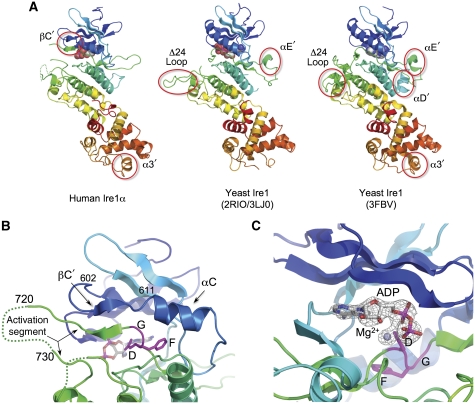
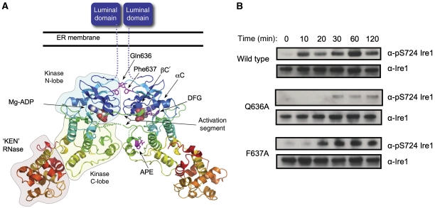
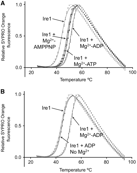
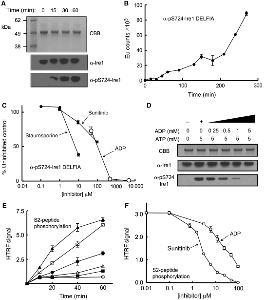
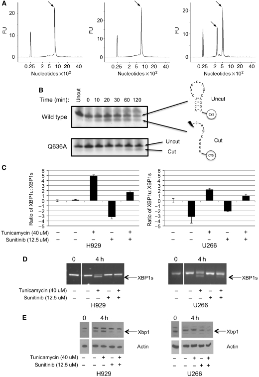
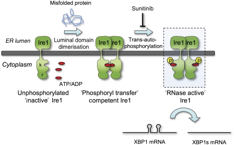
Ire1 (Ern1) is an unusual transmembrane protein kinase essential for the endoplasmic reticulum (ER) unfolded protein response (UPR). Activation of Ire1 by association of its N-terminal ER luminal domains promotes autophosphorylation by its cytoplasmic kinase domain, leading to activation of the C-terminal ribonuclease domain, which splices Xbp1 mRNA generating an active Xbp1s transcriptional activator. We have determined the crystal structure of the cytoplasmic portion of dephosphorylated human Ire1α bound to ADP, revealing the 'phosphoryl-transfer' competent dimeric face-to-face complex, which precedes and is distinct from the back-to-back RNase 'active' conformation described for yeast Ire1. We show that the Xbp1-specific ribonuclease activity depends on autophosphorylation, and that ATP-competitive inhibitors staurosporin and sunitinib, which inhibit autophosphorylation in vitro, also inhibit Xbp1 splicing in vivo. Furthermore, we demonstrate that activated Ire1α is a competent protein kinase, able to phosphorylate a heterologous peptide substrate. These studies identify human Ire1α as a target for development of ATP-competitive inhibitors that will modulate the UPR in human cells, which has particular relevance for myeloma and other secretory malignancies.


Conflict of interest statement

The authors declare that they have no conflict of interest.

Figures

Figure 1
Figure 1
Structure of human Ire1α. (A) Secondary structure cartoons of the crystal structures of cytoplasmic regions of human Ire1α (left) and yeast Ire1 (middle and right), rainbow coloured blue → red, N → C-terminus. Non-canonical secondary structural elements in the yeast structures (αD′, αE′), which mediate protein–protein interactions in the different yeast Ire1 crystal lattices, are not conserved in human Ire1α. Both yeast crystal structures have an engineered loop (Δ24 loop) connecting α-helices EF and F, from which 24 residues were deleted to obtain crystals. The equivalent loop in human (and other metazoan) Ire1α is much shorter, and was not modified. A small helix in the C-terminal RNase domain (α3′), which has been suggested to contribute to the catalytic function of this domain is fully ordered in human Ire1α. Human Ire1α contains an additional β-strand (βC′) formed by the unwinding of the N-terminal end of the kinase domain C-helix, which makes a β-sheet interaction with the N-terminal end of the activation segment. (B) Close-up of the β-sheet interaction between the βC′-strand and the N-terminal part of the activation segment immediately following the conserved DFG motif at residues 711–713. This β-sheet interaction directs the subsequent residues of the activation segment away from the body of the kinase, towards the kinase active site of a second Ire1α molecule (see below). To our knowledge, this type of interaction, involving the unwinding of the C-helix, has not previously been described in protein kinases. The tip of the activation segment between residues 720 and 730 is poorly ordered. (C) Close-up of Mg2+-ADP bound to dephosphorylated human Ire1α. The DFG motif is in the ‘in' conformation associated with an activated kinase, and the side chain of Asp711 in the motif provides a direct ligand interaction with the Mg2+ ion. Electron density mesh is from a Fo–Fc difference Fourier map calculated with Mg-ADP omitted, and contoured at 3.0 σ.
Figure 2
Figure 2
Face-to-face kinase domain dimerisation of human Ire1α. (A) Human Ire1α forms a dimer in which the kinase active sites of the two monomers face each other. The activation segment of one monomer is directed towards the other, so that the target substrate residue, Ser724 would come into close proximity of the Mg2+-ATP bound in the opposite active site, and be phosphorylated by it. This arrangement of Ire1α molecules provides a straightforward mechanistic model for how dimerisation of Ire1 N-terminal domains in the lumen of the ER would facilitate association and transphosphorylation of their associated kinase domains on the cytoplasmic side of the membrane. (B) Autophosphorylation of dephosphorylated dimer interface mutants Q636A and F637A Ire1 as compared with wild type. Both wild-type and mutant proteins were incubated with 5 mM MgCl and 5 mM ATP at 37°C and samples were run at specific time points. Protein samples were visualised by western blot with generic Ire1α or the phospho-specific pS724-Ire1α.
Figure 3
Figure 3
Nucleotide binding. (A) Thermal-shift analysis of adenine nucleotide binding to dephosphorylated Ire1α. ATP and AMPPNP stabilise Ire1 by +4.7°C and +4.1°C, respectively, while ADP shows the tightest binding, stabilising Ire1α by +7.2°C. Reaction conditions are given in Materials and methods section. (B) ADP binding to Ire1α is totally dependent on Mg2+ and in its absence ADP has minimal effect on Ire1α stability.
Figure 4
Figure 4
Autophosphorylation and inhibition. (A) Autophosphorylation of dephosphorylated Ire1α monitored by α-pS724-Ire1α phospho-specific antibody. Ire1α was incubated with 5 mM ATP and 5 mM MgCl2 at 37°C for the times indicated. Protein was visualised by SDS–PAGE with Coomassie Brilliant Blue (CBB) and by western blot with a generic Ire1α antibody or the phospho-specific α-pS724-Ire1α antibody. (B) Quantitative DELFIA assay measuring autophosphorylation of dephosphorylated Ire1α monitored by α-pS724-Ire1α phospho-specific antibody. Reaction used 700 nM Ire1α at 37°C in the presence of 0.5 mM ATP and 20 mM Mg2+. The non-linear kinetics are consistent with a transphosphorylation reaction. (C) Inhibition of Ire1α autophosphorylation by the broad-specificity kinase inhibitor staurosporine, the licensed anti-cancer drug sunitinib, and ADP. Autophosphorylation reaction was followed by DELFIA assay as in (D) after 2.5 h. (D) Inhibition of Ire1α autophosphorylation by ADP. Autophosphorylation of previously dephosphorylated Ire1α, indicated by α-pS724-Ire1α western blot, is progressively inhibited in the presence of increasing concentrations of ADP, and essentially blocked at equimolar ADP to ATP, consistent with the higher affinity of ADP for the nucleotide-binding site in the unphosphorylated kinase indicated by the thermal-shift analysis (A). Reactions were carried out in the presence of 5 mM MgCl2. (E) Hetero-phosphorylation of the HTRF biotinylated peptide S2 (200 nM) by phosphorylated Ire1α (○=no enzyme; ▪=50 nM; ▵=100 nM; •=200 nM; □=400 nM; and ▴=800 nM) in the presence of 30 μM Mg-ATP. (F) Concentration-dependent inhibition of Ire1α hetero-phosphorylation activity. Curves are shown for sunitinib (IC50=3.7 μM±1.2) and ADP (IC50=38 μM±17). Staurosporine (data not plotted) had an IC50=13 nM±8.4. Reactions used 200 nM Ire1α, 200 nM peptide S2, and 30 μM ATP. IC50 values are averages of three experiments.
Figure 5
Figure 5
Xbp1 mRNA cleavage by human Ire1α. (A) Elution profiles from Agilent Bioanalyser of (left) untreated Xbp1 mRNA, (middle) Xbp1 mRNA incubated with dephosphorylated Ire1α, and (right) Xbp1 mRNA incubated with phosphorylated Ire1α. Reaction conditions are given in Materials and methods section. The peak eluting at a position corresponding to 800 nucleotides in the left and middle panels is the unspliced Xbp1 mRNA, whereas the pair of peaks eluting at 300 and 500 nucleotides, respectively, in the right panel, corresponds to the two cleaved products of Xbp1 mRNA. The peak at 25 nucleotides in all three panels is a calibration marker. (B) In vitro cleavage activity of Ire1α, wild-type, and Q636A mutant, on a fluorescently tagged oligonucleotide encapsulating the sequence and secondary structure of a known Ire1α cleavage site in human Xbp1 mRNA. Cleavage generates a fluorescent species with higher gel mobility. The Q636A mutation, which causes a defect in autophosphorylation, results in a similar decrease in RNase activity, confirming the dependence of RNase activation on autophosphorylation. (C) Sunitinib inhibits Xbp1 mRNA splicing in vivo. Myeloma cell lines (U266+H929) were treated with tunicamycin to induce ER stress, in the presence or absence of sunitinib, and levels of spliced (Xbp1s) and unspliced (Xbp1u) mRNAs in the treated cells were determined by quantitative real-time PCR using LUX primers (see Materials and methods), and plotted as relative Xbp1u:Xbp1s ratio. In both cell lines, the addition of the Ire1α kinase inhibitor sunitinib significantly inhibits the splicing of Xbp1 mRNA. (D) Similar results were obtained by RT–PCR amplification. (E) Western blot of protein extracts from cells treated as in (C), showing inhibition of Xbp1 protein production in ER-stressed cells treated with sunitinib. Actin is shown as a loading control.
Figure 6
Figure 6
Autophosphorylation-dependent activation of Ire1α RNase activity. Model of Ire1 dimer-induced mechanism of action, highlighting the role of the face-to-face ‘phosphoryl-transfer' competent state defined here that facilitates transphosphorylation and phosphorylation-dependent downstream activation of the RNase activity. Whether the putatively RNase active back-to-back interaction observed in yeast Ire1 structures also occurs in the mammalian system is yet to be determined—the main structural components, not being conserved—and the mechanism by which autophosphorylation mediates RNase activation is yet to be directly demonstrated. Nonetheless, that inhibition of autophosphorylation by an ATP-competitive inhibitor such as sunitinib blocks consequent RNase activity provides a means by which therapeutic manipulation of the Ire1 arm of the UPRs might be achieved.

References

    1. Adams PD, Gopal K, Grosse-Kunstleve RW, Hung LW, Ioerger TR, McCoy AJ, Moriarty NW, Pai RK, Read RJ, Romo TD, Sacchettini JC, Sauter NK, Storoni LC, Terwilliger TC (2004) Recent developments in the PHENIX software for automated crystallographic structure determination. J Synchrotron Radiat 11: 53–55 - PubMed
    1. Bertolotti A, Zhang Y, Hendershot LM, Harding HP, Ron D (2000) Dynamic interaction of BiP and ER stress transducers in the unfolded-protein response. Nat Cell Biol 2: 326–332 - PubMed
    1. Carrasco DR, Sukhdeo K, Protopopova M, Sinha R, Enos M, Carrasco DE, Zheng M, Mani M, Henderson J, Pinkus GS, Munshi N, Horner J, Ivanova EV, Protopopov A, Anderson KC, Tonon G, DePinho RA (2007) The differentiation and stress response factor XBP-1 drives multiple myeloma pathogenesis. Cancer Cell 11: 349–360 - PMC - PubMed
    1. CCP4 (1994) Programs for protein crystallography. Acta Crystallogr D 50: 760–763 - PubMed
    1. Credle JJ, Finer-Moore JS, Papa FR, Stroud RM, Walter P (2005) On the mechanism of sensing unfolded protein in the endoplasmic reticulum. Proc Natl Acad Sci USA 102: 18773–18784 - PMC - PubMed

Publication types

MeSH terms