. 2011 Apr;18(4):463-70.
doi: 10.1038/nsmb.2018. Epub 2011 Feb 13.

Structural basis for engagement by complement factor H of C3b on a self surface

Affiliations

Structural basis for engagement by complement factor H of C3b on a self surface

Hugh P Morgan et al. Nat Struct Mol Biol. 2011 Apr.

Abstract

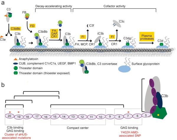
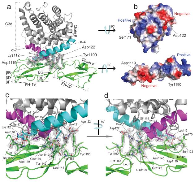
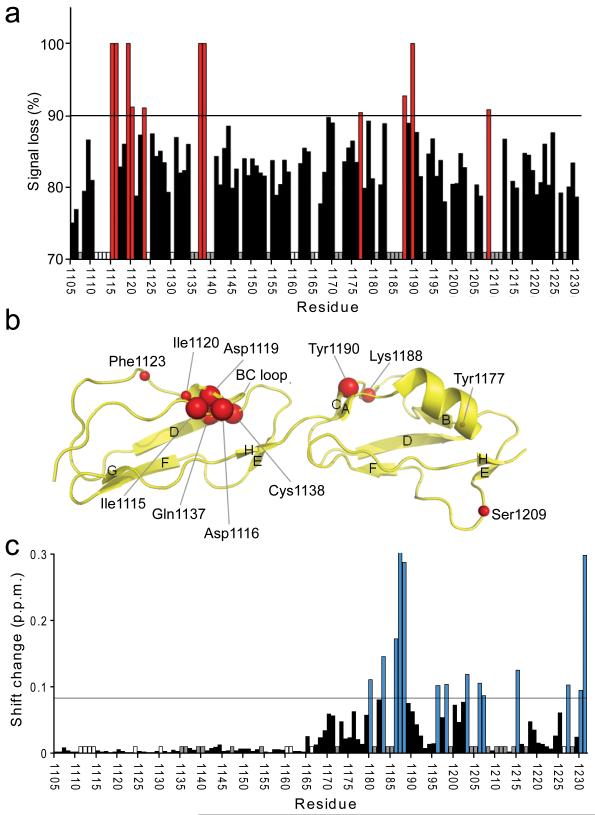
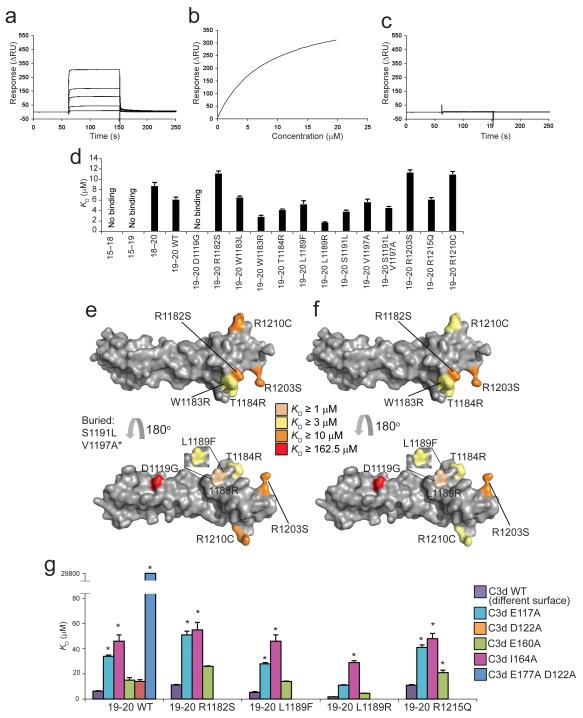
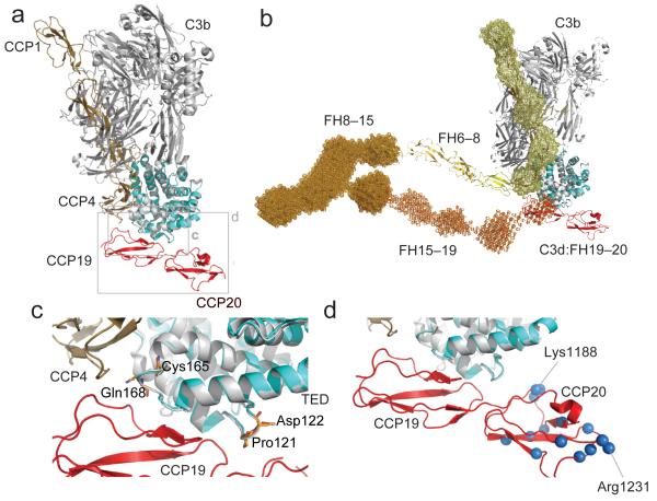
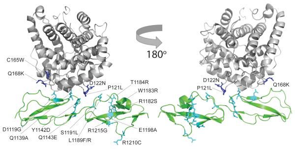
Complement factor H (FH) attenuates C3b molecules tethered by their thioester domains to self surfaces and thereby protects host tissues. Factor H is a cofactor for initial C3b proteolysis that ultimately yields a surface-attached fragment (C3d) corresponding to the thioester domain. We used NMR and X-ray crystallography to study the C3d-FH19-20 complex in atomic detail and identify glycosaminoglycan-binding residues in factor H module 20 of the C3d-FH19-20 complex. Mutagenesis justified the merging of the C3d-FH19-20 structure with an existing C3b-FH1-4 crystal structure. We concatenated the merged structure with the available FH6-8 crystal structure and new SAXS-derived FH1-4, FH8-15 and FH15-19 envelopes. The combined data are consistent with a bent-back factor H molecule that binds through its termini to two sites on one C3b molecule and simultaneously to adjacent polyanionic host-surface markers.


Figures

Figure 1
Figure 1
Introduction to C3b/C3d and complement factor H (FH). (a) The central event in complement activation is C3 cleavage to C3b by C3 convertase accompanied by attachment to surfaces mediated by the TED. In the presence of additional complement regulatory molecules C3b may be further degraded sequentially to iC3b, C3c, C3dg and C3d. C3d corresponds to the TED and remains surface-bound on the cell surface. (b) Complement FH has 20 CCPs and possesses multiple binding sites for different ligands. Disease-linked mutations have been reported throughout FH. Several recombinant fragments of FH were utilized in the current studies and are indicated by black brackets. aHUS - atypical hemolytic uremic syndrome; AMD - age-related macular degeneration.
Figure 2
Figure 2
Structure of the C3d:FH19–20 complex showing interacting domains and overall molecular architecture. All interacting amino acid residues are shown as sticks; H bonds are represented by dashed lines. The resolution of the electron density (2Fo-Fc map) map is 2.1 Å and is contoured at 1σ. (a) Modules 19 (residues 1108–1166) and 20 (residues 1167–1231) of FH are shown in green. Amino acids belonging to two interacting helices (α7 (cyan, residues 170–189) and α4 (purple, residues 106–118) of C3d (shown in grey) form the majority of interactions with FH. (b) Electrostatic surface representations of C3d and FH19–20 (calculated using the APBS plug-in for Pymol 51) rotated 90° counter-clockwise and clockwise, respectively, from the orientations shown in (a). ((c) and (d)) Enlarged views of the C3d and FH19–20 interface.
Figure 3
Figure 3
Heteronuclear NMR spectroscopy used to map the C3d and dp8 binding sites on FH19–20. (a) The percentage broadening observed for unequivocally assigned backbone amides upon addition of C3d is indicated: Red bars signify > 90% broadening; white bars indicate proline residues; and grey bars indicate residues whose assignments were missing or whose intensities were weak in free FH19–20. (b) Amides which experience a line-broadening > 90% are schematically shown as red spheres on the structure of FH19–20 (2G7I). The sizes of the spheres have been adjusted to correlate with the degree of line-broadening that each amide cross-peak experiences: Larger spheres represent complete disappearance of the signal; smaller spheres represent signals broadened by > 90% but which are still detectable. (c) Combined amide chemical shift changes upon addition of 8.5-fold excess of dp8. Blue bars indicate shift changes larger than the threshold (double the average shift change; corresponding residues mapped onto the structure of FH19–20 in Fig. 5d), white and grey bar have the same meaning as in (a).
Figure 4
Figure 4
SPR studies of FH19–20 mutants binding to C3d. (a) Sensorgram showing the binding of wild-type FH19–20 to amine-coupled plasma-derived C3d. (b) KD fitting of (a). (c) Sensorgram showing the very weak binding of FH19–20 D1119G to amine-coupled plasma-derived C3d. (d) Summary of affinity constants of FH fragments and mutants for recombinant C3d. (e) Surface representation of FH19–20 (2G7I) highlighting residues corresponding to mutations that change C3b binding . (f) Surface representation of FH19–20 (2G7I) highlighting residues corresponding to mutations that change C3d binding. Ser1191 and Val1197 are buried residues. *Val1197 has been analyzed in the context of the double mutant S1191L V1197A. Leu1189 has been analyzed in the context of two distinct mutants: L1189R and L1189F. (g) Summary of the KD values for complexes of mutants of FH19–20 and C3d mutants E117A (E1110A), D122A (D1115A) and E160A (E1153A), I164A (I1157A, with wild-type FH19–20 only) and E117A D122A (E1110A D1115A, with wild-type FH19–20 only). The KD of the latter was ~30 mM. * indicates that the corresponding KD value was extrapolated. Error bars (standard error of the mean) for the KD measurements are indicated.
Figure 5
Figure 5
Potential model of FH engagement with surface bound C3b (a) Superposition (C3d on TED) of the C3d:FH19–20 complex and C3b:FH1–4 complex demonstrating the close proximity of FH modules 4 and 19. (b) The FH19–20:C3d structure (red and cyan) is shown superimposed (C3d on TED) onto the FH1–4:C3b structure (tan and gray). This FH model has been constructed by juxtaposition or superposition of the structures of FH5 (yellow) and FH6–8 (yellow-orange) and the SAXS-shape envelopes of FH8-15 (bright orange) and FH15-19 (orange). Structures are shown in cartoon representation; SAXS shape envelopes are shown in mesh. Note, the FH1–4 SAXS shape envelope (pale yellow) is superimposed onto the C3b:FH1–4 structure. (c) Close up view of the α4-α5 loop and the α6-α7 loop of C3d. Highlighted in orange are the residues Pro121 (Pro1092), Asp122 (Asp1093), Cys165 (Cys1136) and Gln168 (Gln1139) for which disease-associated mutations have been reported. (d) Close-up view of the FH19–20 interaction with TED highlighting residues also like to interact with self-surface markers. Amides experiencing considerable chemical shift perturbations upon exposure to dp8 are highlighted as blue spheres (corresponding to their nitrogen atoms). Residues Lys1188 and Arg1231, which exhibit the greatest perturbations upon addition of dp8, are indicated.
Figure 6
Figure 6
The location of mis-sense mutations associated with the development of (aHUS) (and Q1139A) mapped onto the structure of the C3d:FH19–20 complex. The side-chains of C3b mis-sense mutations are shown as blue sticks; side-chains of FH19–20 mis-sense mutations are shown as cyan sticks.

Comment in

  • In self-defense.
    Gros P. Gros P. Nat Struct Mol Biol. 2011 Apr;18(4):401-2. doi: 10.1038/nsmb.2036. Epub 2011 Feb 20. Nat Struct Mol Biol. 2011. PMID: 21336275 No abstract available.

References

    1. Ricklin D, Hajishengallis G, Yang K, Lambris JD. Complement: a key system for immune surveillance and homeostasis. Nat. Immunol. 2010;11:785–97. - PMC - PubMed
    1. de Cordoba SR, de Jorge EG. Translational mini-review series on complement factor H: genetics and disease associations of human complement factor H. Clin. Exp. Immunol. 2008;151:1–13. - PMC - PubMed
    1. Janssen BJ, Christodoulidou A, McCarthy A, Lambris JD, Gros P. Structure of C3b reveals conformational changes that underlie complement activity. Nature. 2006;444:213–6. - PubMed
    1. Pangburn MK, Schreiber RD, Muller-Eberhard HJ. Human complement C3b inactivator: isolation, characterization, and demonstration of an absolute requirement for the serum protein beta1H for cleavage of C3b and C4b in solution. J. Exp. Med. 1977;146:257–70. - PMC - PubMed
    1. Weiler JM, Daha MR, Austen KF, Fearon DT. Control of the amplification convertase of complement by the plasma protein beta1H. Proc. Natl. Acad. Sci. USA. 1976;73:3268–72. - PMC - PubMed

Publication types

Associated data