. 2011 Mar;18(3):262-9.
doi: 10.1038/nsmb.2016. Epub 2011 Feb 13.

Genetic selection designed to stabilize proteins uncovers a chaperone called Spy

Affiliations

Genetic selection designed to stabilize proteins uncovers a chaperone called Spy

Shu Quan et al. Nat Struct Mol Biol. 2011 Mar.

Abstract

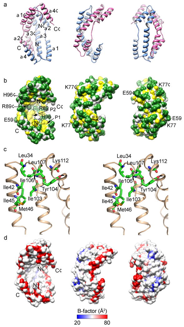
To optimize the in vivo folding of proteins, we linked protein stability to antibiotic resistance, thereby forcing bacteria to effectively fold and stabilize proteins. When we challenged Escherichia coli to stabilize a very unstable periplasmic protein, it massively overproduced a periplasmic protein called Spy, which increases the steady-state levels of a set of unstable protein mutants up to 700-fold. In vitro studies demonstrate that the Spy protein is an effective ATP-independent chaperone that suppresses protein aggregation and aids protein refolding. Our strategy opens up new routes for chaperone discovery and the custom tailoring of the in vivo folding environment. Spy forms thin, apparently flexible cradle-shaped dimers. The structure of Spy is unlike that of any previously solved chaperone, making it the prototypical member of a new class of small chaperones that facilitate protein refolding in the absence of energy cofactors.


Figures

Figure 1
Figure 1
A dual fusion selection for enhancing in vivo protein stability. (a) Unstable test proteins inserted into β-lactamase are degraded by cellular proteases, producing penicillin sensitive (PenVS) strains. Improving the folding of the test proteins increases penicillin resistance (PenVR). (b) Insertion of unstable test proteins into DsbA renders the strains sensitive to cadmium (CdCl2S). Improving the folding of the test proteins increases the strains' resistance towards cadmium (CdCl2R). (c) Thermodynamic stability of Im7 variants (given as ΔΔG0 relative to wild-type Im7) correlates with the minimal inhibitory concentration (MIC) of cadmium for cells expressing the DsbA fusions to these variants. (d) Penicillin resistance of cells expressing the β-lactamase-Im7 fusions correlates with cadmium resistance when they also express the DsbA-Im7 fusion proteins with the same Im7 variants. Error bars indicate the s.d. of three independent measurements.
Figure 2
Figure 2
Overall experimental scheme. Fusion constructs that link the stability of the poorly folded Im7 protein to PenVR (bla∷Im7) and to CdCl2R (dsbA∷Im7) were introduced into the same E. coli strain. Following mutagenesis by EMS treatment, mutants that simultaneously enhanced both PenVR and CdCl2R were selected, and levels of Im7 in these strains were measured. The mutants massively increased levels of Im7 as well as a host protein called Spy. Genetic experiments showed that increased Spy levels are necessary and sufficient to increase Im7 levels. The Spy protein was examined for molecular chaperone activity in vitro and was found to be highly effective as a chaperone in preventing protein aggregation and aiding refolding. Spy was then crystallized, its structure was solved and mutants were made based on its structure to explore the interaction of Spy with its substrates.
Figure 3
Figure 3
Im7 and Spy are abundant in the periplasm of EMS strains. (a) The PenVR and CdCl2R resistant mutants EMS4 and EMS9 and the parental SQ1306 strain were transformed with plasmids encoding wild-type Im7 (WT) or the destabilized variants L53A I54A, I22V, and F84A, . Periplasmic extracts were prepared and analyzed by SDS-PAGE. (b) Spy overexpression is sufficient to enhance Im7 levels in both wild-type and ΔbaeSR backgrounds to those seen in EMS4. Plasmid-encoded Im7 and its destabilized variants were expressed in SQ765 wild-type (WT) or ΔbaeSR backgrounds, with or without the co-expression of plasmid-encoded Spy (designated as Spy++). Aggregated or insoluble Spy is not expected to be extracted using our periplasmic extraction procedure and therefore will not be detected in these experiments.
Figure 4
Figure 4
Spy has chaperone activity. (a) Spy suppresses protein aggregation as monitored by light scattering. Aggregate formation in the absence (0:1) and presence of increasing amounts of Spy (ratios given are Spy:substrate) was monitored for thermally (43 °C) or chemically denatured substrates. (b) Spy enhances protein refolding as assessed by recovery of enzymatic activity. Refolding was monitored in the absence (0:1) and presence of Spy for thermally (43 °C) or chemically denatured substrates. Plots show mean ± s.d. of three independent measurements. Ultracentrifugation and gel filtration studies (Supplementary Fig. 6) indicate that Spy is dimeric in solution, so Spy concentrations are given as a dimer.
Figure 5
Figure 5
Spy protects DsbB, aldolase, and alkaline phosphatase from tannic acid-induced activity loss. Spy concentrations are given as a dimer. Plots show mean ± s.d. for three independent measurements. (a) Enzymatic activity of E. coli DsbB (0.5 μM) incubated in 100 μM tannic acid in the absence (0:1) or presence of increasing amounts of Spy (ratios given are Spy:substrate). (b) Enzymatic activity of rabbit muscle aldolase (0.5 μM) incubated in 16 μM tannic acid in the absence or presence of increasing amounts of Spy. (c) Enzymatic activity of E. coli alkaline phosphatase (AP) (1 μM) incubated in 500 μM tannic acid in the absence or presence of increasing amounts of Spy. (d) Spy and BaeSR deletion strains are tannin sensitive.
Figure 6
Figure 6
Crystal structure of the Spy dimer shown in three orientations rotated by 90° along the vertical axis. For ease of comparison the orientations shown in parts a, b, d are identical. (a) Ribbon drawing shows an all α–helical structure. One subunit is colored light blue and the other is colored magenta. The N and C termini and the secondary structural elements of the molecule are labeled. (b) Surface properties of Spy, colored based on the underlying atoms: backbone atoms, white; polar and charged side-chain atoms, green; hydrophobic side-chain atoms, yellow. Two predominantly hydrophobic patches in the concave surface are indicated as P1 and P2. The lower right patch (P1) is composed of Leu34, Ile42, Met46 and Ile103. The upper left patch (P2) is composed of Pro56, Met64, Ile68, Met85, Met93 and Met97. The residues labeled with fluorescent probes (for experiments in Fig. 7) are circled in black. (c) Stereoview showing the cluster of hydrophobic residues at the tip of the cradle marked as P1 in panel (b). (d) Structural flexibility of Spy dimer. The molecular surface representing the Spy backbone atoms is colored based on the average backbone B-factors for each residue. Note that the rim lining the concave surface has higher B-factors, indicating greater structural flexibility. In particular, the N and C termini are highly mobile. These residues are well conserved among homologous sequences (Supplementary Fig. 5).
Figure 7
Figure 7
Spy binds the disordered model substrate protein casein and the in vivo substrate protein Im7-L53A I54A. (a) Analytical gel filtration of Spy alone, casein alone, or a 1:1 mixture of Spy and casein. The molecular weight of a dimer of Spy is 31 kDa, but it elutes as a 44 kDa molecule, which is consistent with the elongated form of the dimer as seen in the crystal structure. The molecular weight of casein is 23–26 kDa. (b) Competition assay between urea-denatured Im7-L53A I54A and MDH for Spy in MDH refolding. Refolding of chemically denatured MDH was monitored by recovery of enzymatic activity plotted as a fraction of the activity of native (i.e., nondenatured) MDH. Note that the curves of MDH alone (open squares) and MDH + Im7-L53A I54A (closed squares) overlap precisely. (c) Competition assay between casein and MDH for Spy binding. Refolding of chemically denatured MDH was monitored as in (b). (d) Normalized fluorescence emission spectra of acrylodan-labeled Spy mutants H96C and K77C in the absence or presence of an equimolar amount of casein. Note that the fluorescence change upon casein addition for Spy K77-acrylodan is opposite that from Spy H96C-acrylodan. (e) Normalized fluorescence emission spectra of acrylodan-labeled Spy A128C in the absence or presence of an equimolar amount of urea-denatured Im7-L53A I54A.

References

    1. Kerner MJ, et al. Proteome-wide analysis of chaperonin-dependent protein folding in Escherichia coli. Cell. 2005;122:209–220. - PubMed
    1. Duguay AR, Silhavy TJ. Quality control in the bacterial periplasm. Biochim Biophys Acta. 2004;1694:121–134. - PubMed
    1. Foit L, et al. Optimizing protein stability in vivo. Mol Cell. 2009;36:861–871. - PMC - PubMed
    1. Stafford SJ, Humphreys DP, Lund PA. Mutations in dsbA and dsbB, but not dsbC, lead to an enhanced sensitivity of Escherichia coli to Hg2+ and Cd2+ FEMS Microbiol Lett. 1999;174:179–184. - PubMed
    1. De Pascale G, Wright GD. Antibiotic resistance by enzyme inactivation: from mechanisms to solutions. Chembiochem. 2010;11:1325–1334. - PubMed

Publication types

MeSH terms