Comparative Study
. 2010:2010:290516.
doi: 10.1155/2010/290516. Epub 2011 Jan 26.

Green tea epigallocatechin gallate exhibits anticancer effect in human pancreatic carcinoma cells via the inhibition of both focal adhesion kinase and insulin-like growth factor-I receptor

Affiliations
Comparative Study

Green tea epigallocatechin gallate exhibits anticancer effect in human pancreatic carcinoma cells via the inhibition of both focal adhesion kinase and insulin-like growth factor-I receptor

Hoang Anh Vu et al. J Biomed Biotechnol. 2010.

Abstract

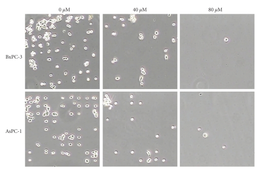
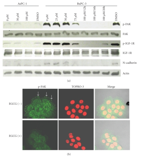
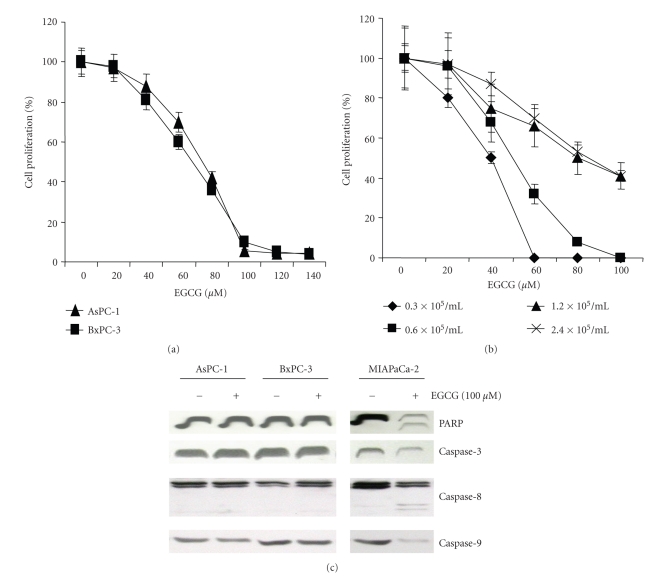
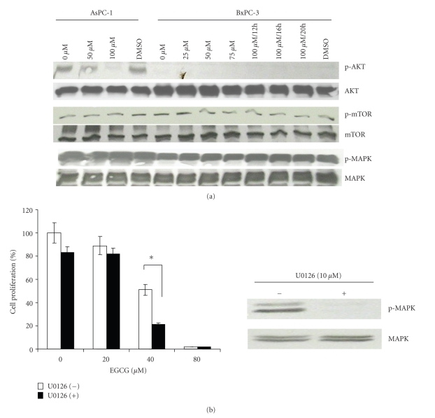
The exact molecular mechanism by which epigallocatechin gallate (EGCG) suppresses human pancreatic cancer cell proliferation is unclear. We show here that EGCG-treated pancreatic cancer cells AsPC-1 and BxPC-3 decrease cell adhesion ability on micro-pattern dots, accompanied by dephosphorylations of both focal adhesion kinase (FAK) and insulin-like growth factor-1 receptor (IGF-1R) whereas retained the activations of mitogen-activated protein kinase and mammalian target of rapamycin. The growth of AsPC-1 and BxPC-3 cells can be significantly suppressed by EGCG treatment alone in a dose-dependent manner. At a dose of 100 μM which completely abolishes activations of FAK and IGF-1R, EGCG suppresses more than 50% of cell proliferation without evidence of apoptosis analyzed by PARP cleavage. Finally, the MEK1/2 inhibitor U0126 enhances growth-suppressive effect of EGCG. Our data suggests that blocking FAK and IGF-1R by EGCG could prove valuable for targeted therapy, which can be used in combination with other therapies, for pancreatic cancer.


Figures

Figure 1
Figure 1
EGCG suppresses cell adhesion by micropattern assay. AsPC-1 and BxPC-3 cells were plated at a density of 0.8 × 105 cells/ml in the presence of indicated doses of EGCG onto micropattern coverslips as described in the Materials and Methods. After 24 hours, cells attaching to single dots were photographed under microscope.
Figure 2
Figure 2
EGCG inhibits FAK activity. (a) Cells were plated at a density of 2 × 10⁵ cells/ml in the presence of various concentrations of EGCG for 12 hours, except for BxPC-3 at 100 μM for additional 4 or 8 hours. Total cell lysates were subjected to western blot analysis with indicated antibodies. Cells cultured in medium with 0.1% DMSO were used as a control. (b) Immunofluorescence staining for p-FAK (green) in BxPC-3 cells in the absence or presence of 100 μM EGCG, as indicated. Expression of p-FAK (arrows) was significantly reduced in EGCG-treated cells as compared with control cells. Staining with TOPRO-3 (red) visualizes the nuclei.
Figure 3
Figure 3
EGCG suppresses cell proliferation. (a) AsPC-1 and BxPC-3 cells at a density of 0.8 × 10⁵ cells/ml were seeded in quadruplicate in 96-well plates, in 100 μl of culture media in the presence of various concentrations of EGCG. After incubation for 48 hours, 10 μl of TetraColor ONE reagent was added to each well, and cells were incubated for additional 4 hours. Absorbance at 450 nm was measured with the Biotrack II plate reader. Results were enumerated as the percentage of the values measured when cells were grown in the absence of EGCG. (b) Cell proliferation assays for BxPC-3 cells at different cell densities. Values were obtained from experiments conducted in triplicate. (c) EGCG fails to cause apoptosis in AsPC-1 and BxPC-3 cells. Cells were plated at a density of 2 × 10⁵ cells/ml in the presence or absence of 100 μM EGCG for 12 hours followed by western blot analysis of total cell lysates using indicated antibodies. MIAPaCa-2 cells were used as a control of apoptosis induced by EGCG.
Figure 4
Figure 4
(a) EGCG fails to inhibit phosphorylation of MAPK and mTOR in AsPC-1 and BxPC-3 cells. Cells were plated at a density of 2 × 10⁵ cells/ml in the presence of various concentrations of EGCG for 12 hours, except for BxPC-3 at 100 μM for additional 4 or 8 hours. Total cell lysates were subjected to western blot analysis. (b) MAPK inhibitor enhances EGCG-induced antiproliferative response in BxPC-3 cells. Cells were plated at a density of 0.6 × 10⁵ cells/ml in 24-well dishes with different concentrations of EGCG for 48 hours in the presence or absence of 10 μM U0126 (MEK1/2 inhibitor). Cells were detached by trypsin, stained with trypan blue and counted manually using a hemacytometer. Values were obtained from experiments conducted in triplicate. Asterisk indicates significant difference. Inhibition of phosphorylation of MAPK by U0126 at 10 μM was confirmed with western blot analysis.

References

    1. Maxwell Parkin D, Bray F, Ferlay J, Pisani P. Estimating the world cancer burden: Globocan 2000. International Journal of Cancer. 2001;94(2):153–156. - PubMed
    1. Hecker TP, Gladson CL. Focal adhesion kinase in cancer. Frontiers in Bioscience. 2003;8:s705–s714. - PubMed
    1. Sieg DJ, Hauck CR, Ilic D, et al. FAK integrates growth-factor and integrin signals to promote cell migration. Nature Cell Biology. 2000;2(5):249–256. - PubMed
    1. Golubovskaya VM, Gross S, Kaur AS, et al. Simultaneous inhibition of focal adhesion kinase and Src enhances detachment and apoptosis in colon cancer cell lines. Molecular Cancer Research. 2003;1(10):755–764. - PubMed
    1. Liu W, Bloom DA, Cance WG, Kurenova EV, Golubovskaya VM, Hochwald SN. FAK and IGF-IR interact to provide survival signals in human pancreatic adenocarcinoma cells. Carcinogenesis. 2008;29(6):1096–1107. - PMC - PubMed

Publication types

MeSH terms