. 2011 Oct;68(19):3233-47.
doi: 10.1007/s00018-011-0630-9. Epub 2011 Feb 12.

Stimulation of olfactory ensheathing cell motility enhances olfactory axon growth

Affiliations

Stimulation of olfactory ensheathing cell motility enhances olfactory axon growth

Louisa C E Windus et al. Cell Mol Life Sci. 2011 Oct.

Abstract

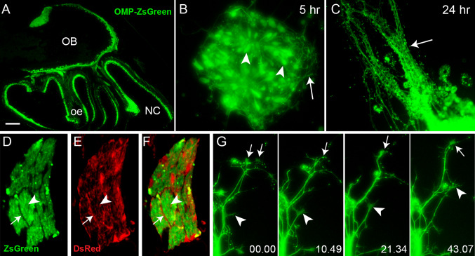
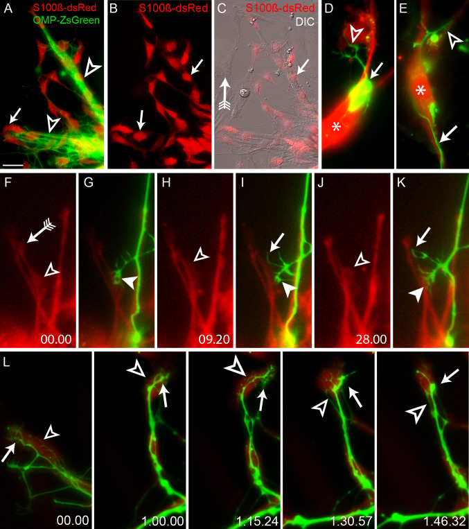
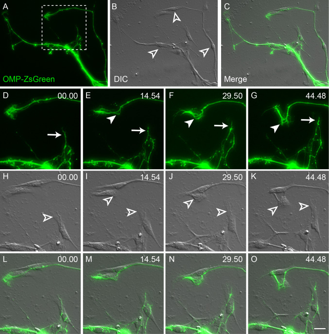
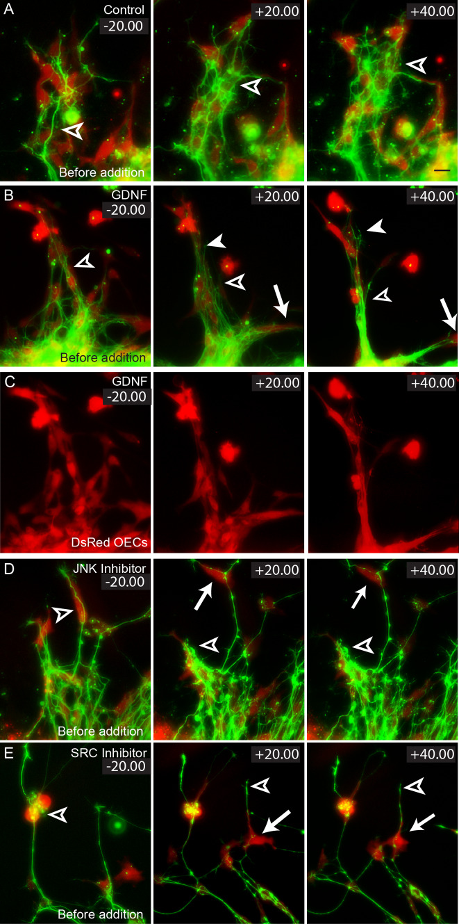
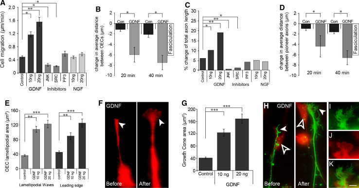
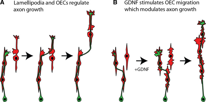
Axons of primary olfactory neurons are intimately associated with olfactory ensheathing cells (OECs) from the olfactory epithelium until the final targeting of axons within the olfactory bulb. However, little is understood about the nature and role of interactions between OECs and axons during development of the olfactory nerve pathway. We have used high resolution time-lapse microscopy to examine the growth and interactions of olfactory axons and OECs in vitro. Transgenic mice expressing fluorescent reporters in primary olfactory axons (OMP-ZsGreen) and ensheathing cells (S100ß-DsRed) enabled us to selectively analyse these cell types in explants of olfactory epithelium. We reveal here that rather than providing only a permissive substrate for axon growth, OECs play an active role in modulating the growth of pioneer olfactory axons. We show that the interactions between OECs and axons were dependent on lamellipodial waves on the shaft of OEC processes. The motility of OECs was mediated by GDNF, which stimulated cell migration and increased the apparent motility of the axons, whereas loss of OECs via laser ablation of the cells inhibited olfactory axon outgrowth. These results demonstrate that the migration of OECs strongly regulates the motility of axons and that stimulation of OEC motility enhances axon extension and growth cone activity.


Figures

Fig. 1a–g
Fig. 1a–g
Olfactory sensory axons extend rapidly in vitro. a P2.5 sagittal section of an OMP-ZsGreen transgenic mouse. Primary olfactory axons express the bright fluorescent protein ZsGreen. b An olfactory epithelium explant prepared from an E12.5 OMP-ZsGreen embryo. After 5 h in culture primary olfactory neurons (arrowheads) were clearly evident and extended nascent axons over the surface of the culture dish (arrow). c After 24 h, axons extended out of the explant and formed tightly fasciculated bundles (arrow). df High magnification view of a cross section through an olfactory sensory axon fascicle; axons (ZsGreen) are ensheathed by olfactory ensheathing cells (DsRed). The OECs line the perimeter (arrow) of fascicles within the large nerve bundle; fine processes (arrowhead) of OECs were also within the fascicle. g Panels show high magnification time-lapse microscopy of ZsGreen-positive axons in culture. Two pioneer axons with active growth cones (arrows 0.00) interacted with each other (arrow 10.49) and fasciculated together (arrow 21.34–43.07) whilst continuing to extend out from the explant. A follower axon extended along the length of the pre-existing axons (arrowhead 0.00). As it extended its active growth cone (arrowhead 10.49), it directly followed and remained in contact with the pre-existing axons (arrowhead 10.49–43.07) via the extension of thin filopodia. Time is recorded in minutes and seconds. OB Olfactory bulb, NC nasal cavity, oe olfactory epithelium. Scale bar 900 μm in a, 10 μm in b, 40 μm in c, 7 μm in df, 20 μm in g
Fig. 2a–l
Fig. 2a–l
Olfactory axon extension is limited by OECs. a A fluorescent ZsGreen image revealed that axons from an explant (out of view at bottom right) extended directly across a chain-like scaffold of OECs (unfilled arrowheads). OECs always migrated ahead of axons (arrow). b The fluorescent DsRed image revealed that OECs migrated out in a chain formation with OECs in direct contact with each other (arrows). c A merged DIC and fluorescent image of the same explant view revealed that DsRed-positive OECs (arrow) migrated out along with cells that were not DsRed-positive (arrow with tail), but that axons always were associated with OECs and not the other cells. d In dissociated cultures of ZsGreen neurons and DsRed OECs, a single neuron (arrow) adhered to the cell body of an OEC (asterisk) and extended a single axon which directly interacted with another OEC (unfilled arrowhead). e An olfactory axon directly adhered to and extended along the shaft of an OEC (arrow), wrapped around the cell body (asterisk) and then extended directly along the shaft of another OEC (unfilled arrowhead). fk Time-lapse imaging revealed that olfactory axon growth cones (GC) formed adhesive contacts with OECs via lamellipodial waves. f OECs exhibited lamellipodial protrusions along their shafts (unfilled arrowhead 00.00) which were independent of the leading edge (arrow with tail). The GC (g filled arrowhead) of a follower axon directly interacted with the lamellipodial wave (f unfilled arrowhead). h After 9.20 min the lamellipodial wave moved along the shaft of the OEC in an anterograde direction (unfilled arrowhead). i A filopodia (arrow) extended from the GC and interacted with the motile lamellipodial wave while the GC remained adhered to the OEC process (arrowhead). j The lamellipodial wave remained active over the course of 28 min (unfilled arrowhead). k The GC (filled arrowhead) remained adhered to the OEC via the lamellipodial wave (j unfilled arrowhead) while the filopodium (arrow) continued to adhere to the OEC process. l Time-lapse microscopy revealed that the extension and motility of pioneer axons mirrored that of OECs. A pioneer axon (arrow) adhered to the shaft of an OEC (unfilled arrowhead). The OEC actively migrated to the right of the field of view (unfilled arrowheads, 00.00–1.15.24) while the axons (arrows) adhered to and responded to the underlying activity of the OEC. The OEC then changed direction (unfilled arrowhead, 1.30.57–1.46.32) and actively migrated while the axon followed (arrows, 1.30.57–1.46.32). Time is recorded in hours, minutes and seconds. Scale bar 40 μm in ac, 10 μm in de, 30 μm in fk, 5 μm in l
Fig. 3a–o
Fig. 3a–o
The motility of olfactory axons is influenced by OEC migration. Panels show axons that have extended out of an E12.5 explant, imaged using ZsGreen fluorescence and differential inference contrast (DIC). a ZsGreen-positive axons extend from the explant which is out of view at the bottom right. b DIC imaging revealed the presence of numerous cells (unfilled arrowheads) underlying the axons. c Merged image of a and b. do Time-lapse imaging of the boxed region shown in a. dg Some ZsGreen axons rapidly extended (arrow) during the imaging period, while others changed direction (filled arrowhead). hk DIC imaging revealed that the underlying cells (unfilled arrowheads) migrated across the surface of the culture dish. lo The ZsGreen-positive axons extended directly along the shaft and body of these cells. At no point did the axons extend out onto the matrix of the culture dish. Time is recorded in minutes and seconds. Scale bar is 15 μm in ac and 3.5 μm in do
Fig. 4a–e
Fig. 4a–e
The effect of GDNF on OECs and olfactory axon extension. Explants of olfactory epithelium were prepared from E14.5 OMP-ZsGreen × S100ß-DsRed embryos, cultured for 24 h and then incubated with either exogenous GDNF (b), JNK (c) or SRC inhibitors (d). ad 20 min prior to challenging OE explants, olfactory axons extended along a chain-like scaffold of DsRed-positive OECs (unfilled arrowheads). a OECs (red) in control explants displayed limited migration and fasciculation during the imaging period. Extension of axons (green) was apparent only with follower axons (unfilled arrowheads). b After the addition of GDNF, OECs displayed higher migration rates (arrows, +20.00, +40.00) and formed tightly fasciculated bundles (unfilled arrowheads). Pioneer olfactory axons clearly exhibit additional extension and fasciculation (filled arrowhead). c DsRed-only fluorescence of images shown in b shows the changes in OEC distribution. d With the addition of JNK inhibitor, OEC migration (arrows, +20.00, +40.00) and axon extension (unfilled arrowheads) were minimal. e Similarly with the addition of SRC inhibitor, OEC migration (arrows, +20.00, +40.00) and axon extension (unfilled arrowheads) were minimal. Time is recorded in minutes and seconds. Scale bar 20 μm
Fig. 5a–k
Fig. 5a–k
GDNF influences OEC migration and pioneer axon extension. a OECs migrated at higher rates with addition of 10 and 20 ng/ml of GDNF; the addition of JNK (9SP600125) or SRC (PP2) inhibitors decreased migration of OECs; incubation with the inactive analogue PP3 or NGF had no effect on OEC migration (n = 35–40; p < 0.01, Kruskal-Wallis test and *p < 0.05, **p < 0.01, post hoc Dunn’s multiple comparison test). b Quantification of the average distance between OECs when incubated with GDNF (n = 10; p < 0.05, Kruskal-Wallis test and *p < 0.05, post hoc Dunn’s multiple comparison test). c Quantification of pioneer axon extension. Axons extended at higher rates when incubated with 10 and 20 ng/ml of GDNF; the addition of JNK (9SP600125) or SRC (PP2) inhibitors decreased axon extension; incubation with the inactive analogue PP3 or NGF had no effect on axon extension (n = 19–20; p < 0.01, Kruskal-Wallis test and *p < 0.05, **p < 0.01, post hoc Dunn’s multiple comparison test). d Quantification of the average distance between pioneer axons when incubated with GDNF (n = 10; p < 0.05, Kruskal-Wallis test and *p < 0.05, post hoc Dunn’s multiple comparison test. e Quantification of the surface area of lamellipodial waves and leading edge on OECs incubated with GDNF (n = 40–45; p < 0.001, Kruskal-Wallis test and **p < 0.01, ***p < 0.001, post hoc Dunn’s multiple comparison test). f Addition of GDNF dramatically increased the size of the leading edge on OECs. g Quantification of the surface area of growth cones (GC) on axons incubated with GDNF (n = 38–45; p < 0.001, Kruskal-Wallis test and ***p < 0.001, post hoc Dunn’s multiple comparison test). h Addition of GDNF dramatically increased the size of the GC (filled arrowhead) on axons; example shown is one of the more dramatic increases in GC area. An underlying OEC (unfilled arrowhead) migrated during the assay. i There was a direct relationship between increased size in GC on axons and increased size of lamellipodia on OECs (j); GC size would increase when in direct contact with OEC lamellipodia (k). Error bars denote SEM. Scale bar 10 μm in f, hk
Fig. 6a–i
Fig. 6a–i
Ablation of pre-existing OEC scaffolds inhibits olfactory axon extension. Panels ac show fluorescence and DIC combined images; panels df show same images but without DIC. a, d Pre-ablation: DsRed-positive OECs migrated out of an explant onto the matrix forming a chain-like scaffold (unfilled arrowheads). ZsGreen-positive axons (arrows) extended directly across the OEC scaffold. b, e Two hours post ablation: DsRed-positive OECs were no longer present on the matrix (unfilled arrowheads) but remained active in the non-ablated region of the explant (black arrowheads). Olfactory axons remained active across the matrix (arrows). c Eight hours post ablation: very few olfactory axons remained within the ablated region (arrow) despite the presence of other cell types migrating across the matrix (black arrowhead). f DsRed-positive OECs started to migrate out of the explant (unfilled arrowhead) accompanied by the regrowth of olfactory axons (arrow). g For the control, a different region of the same explant was used. h OECs in the control region were not ablated and robust axon outgrowth occurred. Scale bar 20 μm in af; 40 μm in gh. i Quantification of the change in axon length after ablating pre-existing OEC scaffolds. The length of olfactory axons significantly declined at 2 and 8 h post ablation compared to control axons grown on OECs of the same explants (n = 7; p < 0.01, Kruskal-Wallis test and **p < 0.01, post hoc Dunn’s multiple comparison test)
Fig. 7
Fig. 7
a Olfactory axons (green) extend along the surface of OECs (red). Growth cones contact lamellipodial waves on OECs and then further extend along the new OEC. The migration of the OECs strongly regulates the motility of the axon. b GDNF stimulates the lamellipodial waves on OECs which results in close adhesion and contact-mediated migration of the OECs. The axons and their growth cones mirror the movement of the OECs

References

    1. Doucette JR. Glial influences on axonal growth in the primary olfactory system. Glia. 1990;3:433–449. doi: 10.1002/glia.440030602. - DOI - PubMed
    1. Franceschini I, Barnett S. Low-affinity NGF-receptor and E-N-CAM expression define two types of olfactory nerve ensheathing cells that share a common lineage. Dev Biol. 1996;173:327–343. doi: 10.1006/dbio.1996.0027. - DOI - PubMed
    1. Doucette JR. Development of the nerve fiber layer in the olfactory bulb of mouse embryos. J Comp Neurol. 1989;285:514–527. doi: 10.1002/cne.902850407. - DOI - PubMed
    1. Chuah MI, Choi-Lundberg D, Weston S, Vincent AJ, Chung RS, Vickers JC, West AK. Olfactory ensheathing cells promote collateral axonal branching in the injured adult rat spinal cord. Exp Neurol. 2004;185:15–25. doi: 10.1016/j.expneurol.2003.09.008. - DOI - PubMed
    1. Chung RS, Woodhouse A, Fung S, Dickson TC, West AK, Vickers JC, Chuah MI. Olfactory ensheathing cells promote neurite sprouting of injured axons in vitro by direct cellular contact and secretion of soluble factors. Cell Mol Life Sci. 2004;61:1238–1245. doi: 10.1007/s00018-004-4026-y. - DOI - PMC - PubMed

Publication types

Substances

LinkOut - more resources