. 2012 Feb;34(1):27-41.
doi: 10.1007/s11357-011-9211-y. Epub 2011 Feb 12.

Sequential stages in the age-dependent gradual formation and accumulation of tubular aggregates in fast twitch muscle fibers: SERCA and calsequestrin involvement

Affiliations

Sequential stages in the age-dependent gradual formation and accumulation of tubular aggregates in fast twitch muscle fibers: SERCA and calsequestrin involvement

Simona Boncompagni et al. Age (Dordr). 2012 Feb.

Abstract

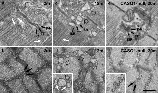
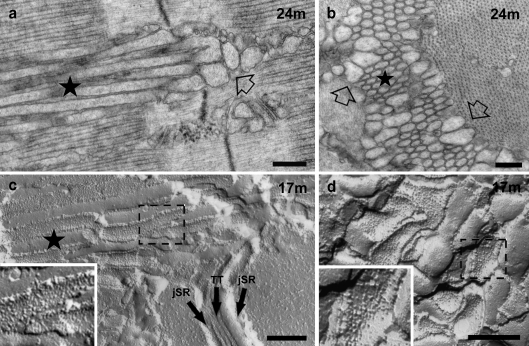
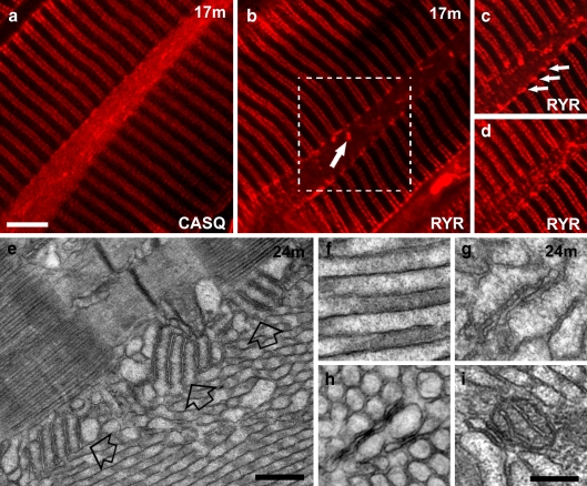
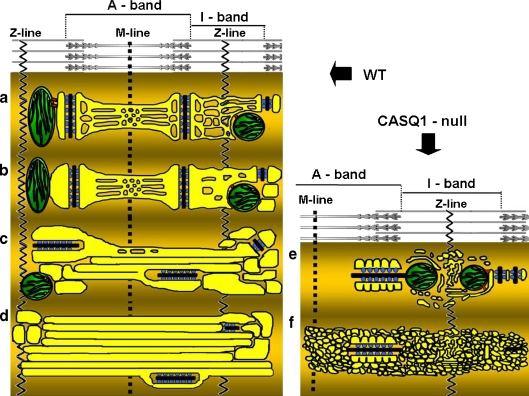
Tubular aggregates (TAs), ordered arrays of elongated sarcoplasmic reticulum (SR) tubules, are present in skeletal muscle from patients with myopathies and are also experimentally induced by extreme anoxia. In wild-type mice TAs develop in a clear age-, sex- (male), and fiber type- (fast twitch) dependence. However, the events preceding the appearance of TAs have not been explored. We investigated the sequential stages leading to the initial appearance and maturation of TAs in EDL from male mice. TAs' formation requires two temporally distinct steps that operate via different mechanisms. Initially (before 1 year of age), the SR Ca(2+) binding protein calsequestrin (CASQ) accumulates specifically at the I band level causing swelling of free SR cisternae. In the second stage, the enlarged SR sacs at the I band level extend into multiple, longitudinally oriented tubules with a full complement of sarco(endo)plasmic reticulum Ca(2+) ATPases (SERCA) in the membrane and CASQ in the lumen. Tubules gradually acquire a regular cylindrical shape and uniform size apparently in concert with partial crystallization of SERCA. Multiple, small TAs associate to form fewer mature TAs of very large size. Interestingly, in fibers from CASQ1-knockout mice abnormal aggregates of SR tubules have different conformation and never develop into ordered aggregates of straight cylinders, possibly due to lack of CASQ accumulation. We conclude that TAs do not arise abruptly but are the final result of a gradually changing SR architecture and we suggest that the crystalline ATPase within the aggregates may be inactive.


Figures

Fig. 1
Fig. 1
The Initial step in the formation of TAs is the remodeling of the SR at the I band in IIB/IIX fibers of male mice. a and b SR is constituted of jSR (labeled), forming triads with T-tubules (TT) and free SR, composed of narrow, meandering tubules (a, empty and white arrow). At the I band level (b, black arrows) the free SR is arranged in two to three layers. At 2 to 3 months of age (a, b) the I band free SR has small, mostly empty profiles. c, d At 12 months, both jSR (labeled) and free-SR-sectioned profiles at the I band are often wider and have a visible electron dense content (empty arrows). Dilation of the free SR does not occur at the A band level (c, white arrow). The enlarged detail in d shows medium (white arrows) and large (empty arrow) SR profiles. e, f In CASQ1-null EDL fibers, the jSR cisternae (labeled) are reduced in size compared to WT. Neither jSR nor free SR change size and shape with increasing age in absence of CASQ1 (f, arrows). jSR junctional SR; TT transverse tubules. Bar 0.5 μm
Fig. 2
Fig. 2
The second step in the formation of TAs is the elongation and aggregation of parallel SR tubules, which protrude from the I into the A band. a The dilated SR tubules at the I band (empty arrow) elongate with age into longitudinal tubules that aggregate into parallel bundles (nascent TA, star). These bundles of tubules invade intermyofibrillar spaces at all levels of the sarcomere. b TAs are constituted of a peripheral ring of dilated SR profiles and irregularly shaped tubules (empty arrows) surrounding a central core of uniformly sized and regularly spaced tubules (star). c, d Parallel stacks of tubules growing from the I band into the A band are also visible in FF replicas (nascent TA, star in c). TA tubules (c) and dilated SR at the I band (d) are both uniformly covered by particles representing SERCA (dashed boxes). Bars 0.25 μm
Fig. 3
Fig. 3
TAs become larger and more frequent with age. a Phase contrast image showing two elongated striation free islands marking the position of TAs (stars). b, c Large TAs result from close association of multiple TAs, each with its ring of wider, more irregular tubules (c, empty arrow) and a central core of parallel and uniformly sized tubules. d, e The perfectly regular and straight shape of the core tubules is emphasized by freeze fracture replicas, where they appear as regular cylinders (d, star). The tubules transversely fractured in e displaying regular diameters. Barsa 20 μm; b 0.5 μm; c 2 μm; d 1 μm; e 0.25 μm
Fig. 4
Fig. 4
In the core of large TAs, SR tubules are regularly arranged, perfectly cylindrical, covered by SERCA particles, and held together by little bridges. a, b Tubules in the center of TAs are arranged in a highly ordered hexagonal pattern (a), giving rise to an electron optical diffraction transform that indexes on 60° angles and shows up to five orders of reflections (b). c The ordered arrangements of tubules is probably maintained, if not established, by frequent cross links of uniform size that bridge the gap between the membrane of adjacent tubules (small arrows). d Tubules in the center of large TAs are perfectly straight resulting—when freeze-fractured and replicated—in regularly alternated views of the luminal and cytoplasmic (large and small arrows, respectively) leaflets. e A diagram modeling the plane of fracture that exposes alternatively luminal and cytoplasmic leaflets of the tubules (large and small arrows, respectively). f The cytoplasmic leaflet is decorated by particles representing small aggregates of SERCA molecules. The particles show tendency towards a helical arrangement (f, arrows), which is best detected by holding the image at eye level and glancing along the direction of the two sets of arrows. g A more precise view of the helical symmetry of SERCA particles (arrows) is visible on the free cytoplasmic surface of an SR tubule isolated from aging muscle and prepared by freeze-drying, rotary shadowing. Barsa, d 0.1 μm; b, e, g 0.05 μm
Fig. 5
Fig. 5
TAs contain CASQ, but not RyRs. Mitochondria, T-tubules, and triads are only occasionally trapped within the aggregate. a, b Immunostaining of EDL fibers with antibodies against CASQ and RYR reveals that TAs contain CASQ (a, see also f), but not RYR (b). Bright RYR-positive spots (arrow in b), representing trapped triads (see also g), are rare. c, d Two different optical sections of the area marked in b by the dashed box show RYR positive spots at the edges of a optically sectioned TA (c, arrows; see also e) and in a plane grazing the TA (d). e Stacks of SR-T-tubules junctions (empty arrows) containing RYRs are frequently present at the TA’s edge (compare with c). f TAs’ tubules are filled with electron-dense material, most likely CASQ. g–i SR/T-tubule junctions (or triads, g), T-tubules (h), and mitochondria (i) are rarely trapped inside TAs, mostly where smaller TAs merge with each other (see also Fig. 3b). Barsad, 5 μm; e 0.5 μm; fi 0.2 μm
Fig. 6
Fig. 6
Abnormal aggregates of SR tubules in CASQ1-null fibers are present, but they never became straight, cylinder shaped, and orderly. a, b CASQ1-null aggregates of SR tubules (a, star), continuous with the jSR of triads (a, b, arrows), are composed of tubules/sacs smaller in diameter that those described for TAs in WT fibers and remain convoluted. The jSR cisternae in these fibers are periodically pinched (b, arrowheads) and display a scalloped outline. c Characteristic stacks of flat SR cisternae (empty arrow) are occasionally present immediately next to SR aggregates. These stacks are tethered together by small bridges (detail in c, small arrows). d In freeze-fracture replicas, the CASQ1-null SR aggregates contain particles, presumably SERCA, in the cytoplasmic leaflet, which do not show any sign of helical symmetry (detail in d). Barsa 0.5 μm; bd 0.2 μm
Fig. 7
Fig. 7
Modeling of ad sequential steps leading to formation of TAs in WT fast glycolytic fibers; e, f impaired assembly of TAs in CASQ1-null fibers. a Normal morphology of SR and triads in adult fibers. In adult mammalian muscle, the SR (yellow) is comprised of two domains: (1) jSR constituting the two lateral sacs of a triads, comprising two arrays of RYRs (blue); and (2) free SR, that extends on both sides of the triad, along the entire sarcomere. The T tubule is in black. The I band free SR is composed mostly of irregular and convoluted tubules, whereas the A band free SR is a combination of tubules and flat fenestrated cisternae (see Fig. 1). b Dilatation of I band SR. Around 12 months of age, junctional and free SR at the I band level of fast twitch fibers in males is often abnormally dilated, whereas A band free SR maintains its usual shape (see Fig. 1). c Elongation of tubules into the A band. With increasing age (17–24 months) the dilated junctional and free I band SR projects elongated, cylindrical longitudinal tubules. Triads become more frequently longitudinal (see Fig. 2). d Formation of large tubular aggregates. Growing accumulation of tubules becomes TAs that displace all other cell organelles and coalesce into increasingly larger aggregates. Dilated SR cisternae and triads are mostly confined to the TAs edges (see Figs. 3 and 5). e, f TAs do not develop without CASQ1. In CASQ1-null fibers the jSR cisternae width is significantly reduced compared to the WT (Paolini et al. 2007). The membrane of jSR cisternae displays a scalloped profile on the side opposite to the T-tubule.CASQ1-null SR tubules at the I band level remains narrow and very convoluted, even where they occasionally accumulate into aggregates reminiscent of but not identical to TAs

References

    1. Agbulut O, Destombes J, Thiesson D, Butler-Browne G. Age-related appearance of tubular aggregates in the skeletal muscle of almost all male inbred mice. Histochem Cell Biol. 2000;114(6):477–481. - PubMed
    1. Airey JA, Beck CF, Murakami K, Tanksley SJ, Deerinck TJ, Ellisman MH, Sutko JL (1990) Identification and localization of two triad junctional foot protein isoforms in mature avian fast-twitch skeletal muscle. J Biol Chem 265:14187–14194 - PubMed
    1. Augusto V, Padovani CR, Campos GER. Skeletal muscle fiber types in c57bl6j mice. Braz J morphol Sci. 2004;21(2):89–94.
    1. Boncompagni S, d’Amelio L, Fulle S, Fano G, Protasi F. Progressive disorganization of the excitation-contraction coupling apparatus in aging human skeletal muscle as revealed by electron microscopy: A possible role in the decline of muscle performance. J Gerontol A Biol Sci Med Sci. 2006;61(10):995–1008. doi: 10.1093/gerona/61.10.995. - DOI - PubMed
    1. Boncompagni S, Loy RE, Dirksen RT, Franzini-Armstrong C. The I4895T mutation in the type 1 ryanodine receptor induces fiber-type specific alterations in skeletal muscle that mimic premature aging. Aging Cell. 2010;9(6):958–970. doi: 10.1111/j.1474-9726.2010.00623.x. - DOI - PMC - PubMed

Publication types

Substances