

Coordination features and affinity of the Cu²+ site in the α-synuclein protein of Parkinson's disease
- PMID: 21319811
- PMCID: PMC3064431
- DOI: 10.1021/bi101912q
Coordination features and affinity of the Cu²+ site in the α-synuclein protein of Parkinson's disease
Abstract
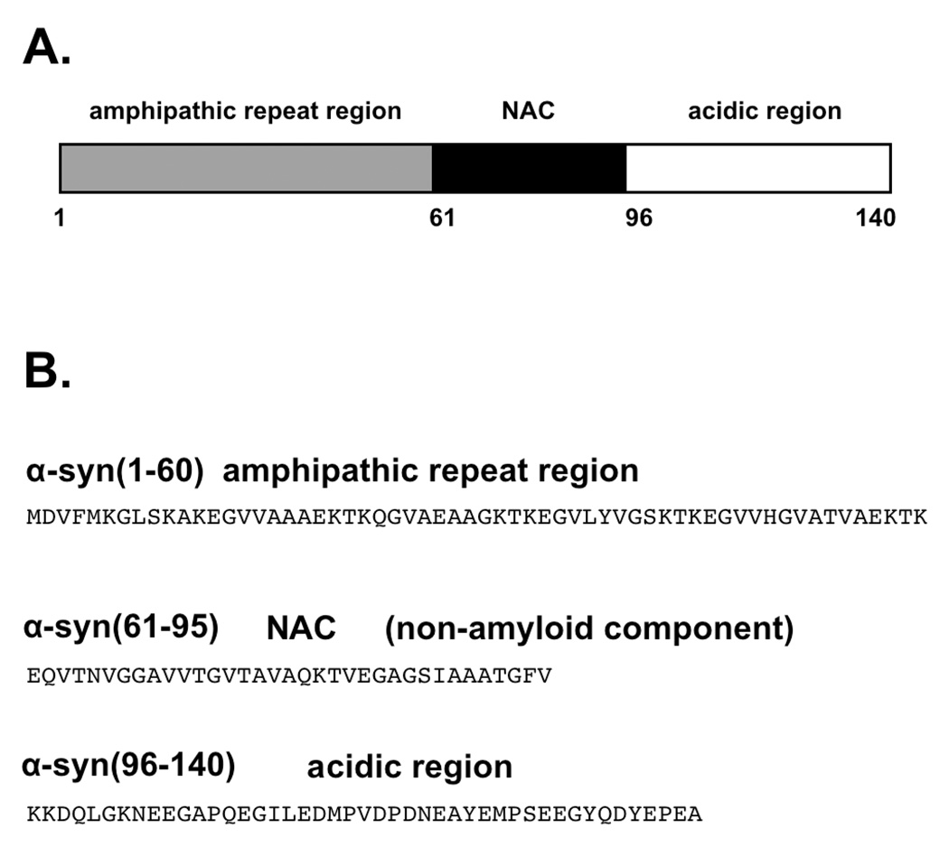
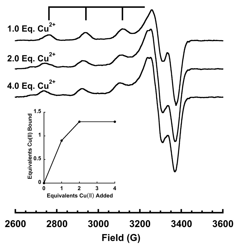
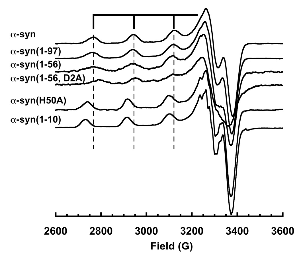
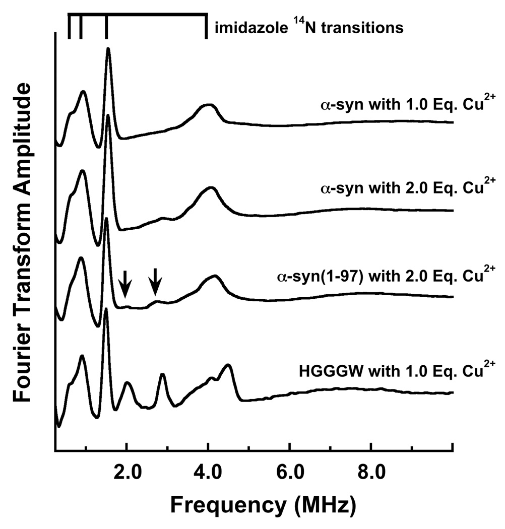
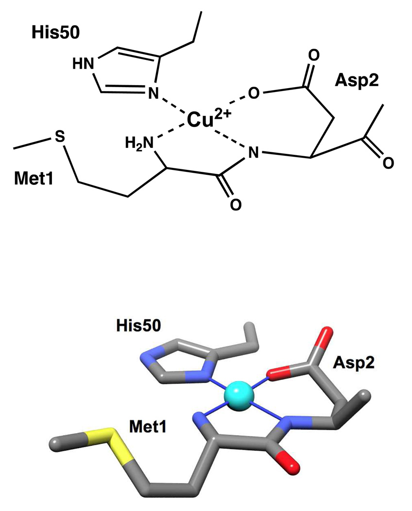
Parkinson's disease (PD) is the second most prevalent age-related, neurodegenerative disorder, affecting >1% of the population over the age of 60. PD pathology is marked by intracellular inclusions composed primarily of the protein α-synuclein (α-syn). These inclusions also contain copper, and the interaction of Cu(2+) with α-syn may play an important role in PD fibrillogenesis. Here we report the stoichiometry, affinity, and coordination structure of the Cu(2+)-α-syn complex. Electron paramagnetic resonance (EPR) titrations show that monomeric α-syn binds 1.0 equiv of Cu(2+) at the protein N-terminus. Next, an EPR competition technique demonstrates that α-syn binds Cu(2+) with a K(d) of ≈0.10 nM. Finally, EPR and electron spin echo modulation (ESEEM) applied to a suite of mutant and truncated α-syn constructs reveal a coordination sphere arising from the N-terminal amine, the Asp2 amide backbone and side chain carboxyl group, and the His50 imidazole. The high binding affinity identified here, in accord with previous measurements, suggests that copper uptake and sequestration may be a part of α-syn's natural function, perhaps modulating copper's redox properties. The findings further suggest that the long-range interaction between the N-terminus and His50 may have a weakening effect on the interaction of α-syn with lipid membranes, thereby mobilizing monomeric α-syn and hastening fibrillogenesis.
Figures
References
-
- Lees AJ, Hardy J, Revesz T. Parkinson's disease. Lancet. 2009;373:2055–2066. - PubMed
-
- Jankovic J. Parkinson's disease: clinical features and diagnosis. J Neurol Neurosurg Psychiatry. 2008;79:368–376. - PubMed
-
- Chartier-Harlin MC, Kachergus J, Roumier C, Mouroux V, Douay X, Lincoln S, Levecque C, Larvor L, Andrieux J, Hulihan M, Waucquier N, Defebvre L, Amouyel P, Farrer M, Destee A. Alpha-synuclein locus duplication as a cause of familial Parkinson's disease. Lancet. 2004;364:1167–1169. - PubMed
-
- Vila M, Przedborski S. Genetic clues to the pathogenesis of Parkinson's disease. Nat Med. 2004;10 Suppl:S58–S62. - PubMed
Publication types
MeSH terms
Substances
Grants and funding
LinkOut - more resources
Full Text Sources
Other Literature Sources
Molecular Biology Databases
Miscellaneous
