. 2011 May 1;435(3):711-22.
doi: 10.1042/BJ20101726.

Molecular interaction and functional regulation of connexin50 gap junctions by calmodulin

Affiliations

Molecular interaction and functional regulation of connexin50 gap junctions by calmodulin

Yanyi Chen et al. Biochem J. .

Abstract

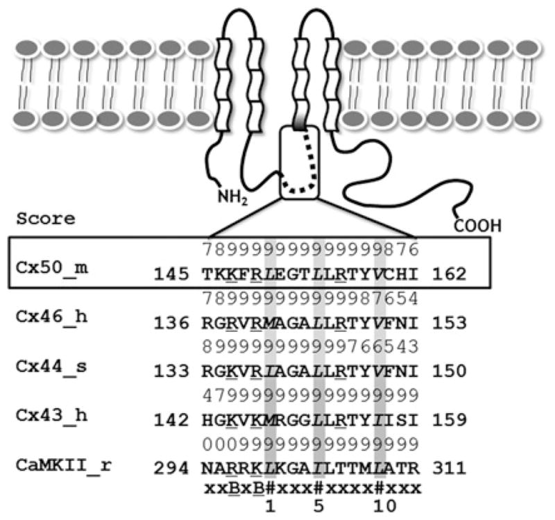
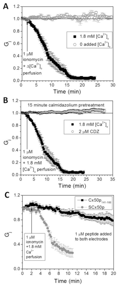
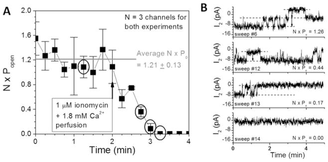
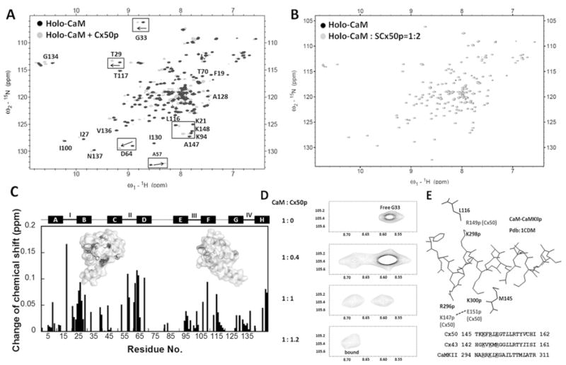
Cx50 (connexin50), a member of the α-family of gap junction proteins expressed in the lens of the eye, has been shown to be essential for normal lens development. In the present study, we identified a CaMBD [CaM (calmodulin)-binding domain] (residues 141-166) in the intracellular loop of Cx50. Elevations in intracellular Ca2+ concentration effected a 95% decline in gj (junctional conductance) of Cx50 in N2a cells that is likely to be mediated by CaM, because inclusion of the CaM inhibitor calmidazolium prevented this Ca2+-dependent decrease in gj. The direct involvement of the Cx50 CaMBD in this Ca2+/CaM-dependent regulation was demonstrated further by the inclusion of a synthetic peptide encompassing the CaMBD in both whole-cell patch pipettes, which effectively prevented the intracellular Ca2+-dependent decline in gj. Biophysical studies using NMR and fluorescence spectroscopy reveal further that the peptide stoichiometrically binds to Ca2+/CaM with an affinity of ~5 nM. The binding of the peptide expanded the Ca2+-sensing range of CaM by increasing the Ca2+ affinity of the C-lobe of CaM, while decreasing the Ca2+ affinity of the N-lobe of CaM. Overall, these results demonstrate that the binding of Ca2+/CaM to the intracellular loop of Cx50 is critical for mediating the Ca2+-dependent inhibition of Cx50 gap junctions in the lens of the eye.


Figures

Figure 1
Figure 1. Cxs membrane topology and the putative CaM-binding sites
The α-class of Cxs are composed of four transmembrane segments, two extracellular loops, one cytoplasmic loop, a short N-terminus and a much longer C-terminal tail. The predicted CaM-binding sites are located in the second half of the intracellular loop between TM2 and TM3. The numeric score (1–9) represents the probability of an accurate prediction of high-affinity CaM-binding sites. The CaM-binding sequences identified in the α-class Cxs are similar to those of CaMKII. All of the aligned sequences fit the 1–5–10 CaM-binding mode subclass, where each number represents the presence of a hydrophobic residue. m, mouse; h, human; s, sheep; r, rat; #, hydrophobic residues (highlighted in grey); B, basic residues (underscored).
Figure 2
Figure 2. Ca2+- and CaM-dependence of Cx50 gap junction uncoupling
(A) Cx50-N2a cell pair superfusion with 1 μM ionomycin bath saline with or without 1.8 mM CaCl2 demonstrates a time-dependent uncoupling of Cx50 gap junctions that requires 1.8 mM [Ca2+]o. (B) Pre-treatment with 2 mM CDZ for 10–15 min before patch-clamp analysis completely prevented the 1.8 mM [Ca2+]o-dependent uncoupling response. (C) Inclusion of 1 μM Cx50p141–166 in both whole-cell patch pipettes prevented 80 % of the Ca2+/CaM-dependent decline in Cx50 Gj. In contrast, experiments performed with SCx50p prevented only 20 % of the 1.8 mM [Ca2+]o–CaM-dependent reduction in Cx50 Gj. The average ± S.E.M. initial gj values for all experimental groups are displayed in Table 1.
Figure 3
Figure 3. Mechanistic basis for Ca2+/CaM-dependent Cx50 gap junction uncoupling
(A) The product of the number of open channels (N) and open probability (P o) from two poorly coupled Cx50-N2a cell pairs is plotted as a function of time and 1 μM ionomycin +1.8 mM [Ca2+]o superfusion. N · Po declined from an average control value of 1.21 open channels to 0 within 2 min of ionomycin/CaCl2 saline superfusion. (B) Cx50 gap junction channel current recordings from one experiment are shown for the time points indicated by the circles in (A). The number and duration of the open channels declined progressively to 0 without any apparent reduction in the single-channel conductance (314 pS), indicative of closure of a Ca2+–CaM-dependent gate without a block of the ion-permeation pathway.
Figure 4
Figure 4. Monitoring the interaction between CaM and Cx50p141–166 by (1H,15N)-HSQC spectroscopy
(A) An overlay of HSQC spectra of holo-CaM (black) with the spectrum of the holo-CaM–Cx50p141–166 (grey). (B) An overlay of the HSQC spectra of holo-CaM (black) with the spectrum of the holo-CaM–SCx50p (grey). (C) Chemical-shift perturbation in CaM induced by addition of 2-fold molar excess of Cx50p141–166. The weight-average chemical-shift change (Δδ) was calculated using eqn (5). Residues with Δδ> 0.1 p.p.m. were mapped to the three-dimensional structure of holo-CaM (PDB code 3CLN). (D) The chemical-shift change of Gly33 during titration of holo-CaM with Cx50p141–166. The disappearance of the peak (free form) was accompanied by the appearance of the corresponding peak (bound form) at a new position. (E) Structural basis of the difference in directionality of chemical shift change. The structure of the CaM–CaMKII complex (PDB code 1CDM) is shown with the peptide (inside) and the CaM residues (outside). Leu116 and Met145 of CaM are within 5 Å of Lys298p and Lys300p of the peptide respectively. These peptide positions correspond to Lys146p and Arg148p for Cx43, and Arg149p and Glu151p for Cx50, as can be seen in the superposition of their sequences. In Cx50, Glu151p may be pulled away from Met145 by Lys147p. In contrast, in Cx43, Arg148p may be pushed towards Met145 by Lys144p. These differences in peptide sequences can cause changes of chemical shifts in different directions.
Figure 5
Figure 5. Hydrodynamic radii of CaM–Cx50p141–166 complex determination by pulse-field gradient NMR
The NMR signal decay of holo-CaM (○) and holo-CaM–Cx50p141–166 complex (●) as a function of field strength. The calculated hydrodynamic radii of the CaM and complexes are indicated on the top.
Figure 6
Figure 6. CD studies of the interaction between CaM and Cx50p141 – 166
(A) Far-UV CD spectra of CaM in the presence of 5 mM EGTA (○, apo-CaM) or CaCl2 (□, holo-CaM), and a CaM–Cx50p141–166 (1:1) mixture with 5 mM EGTA (●, apo-CaM–Cx50p141–166) or CaCl2 (■, holo-CaM–Cx50p141–166). (B) Far-UV spectra of the synthetic peptide Cx50p141–166 with addition of TFE (0–80 %). The inset shows calculated α-helical content as a function of TFE concentration for the peptides Cx50p141–166 (■), Cx44p129–150 (▲) and Cx43p136–158 (●). (C) Far-UV spectra of the synthetic peptide Cx50p141–166 with (●) or without (○) addition of CaM.
Figure 7
Figure 7. Interaction of Cx50p141–166 with D-CaM monitored by steady-state fluorescence
(A) The titration curve of D-CaM (2 μM) with Cx50p141–166 in the presence of 5 mM Ca2+ in a buffer consisting of 50 mM Tris/HCl (pH 7.5) and 100 mM KCl. The inset shows the fluorescence spectrum of D-CaM in the absence (broken line) or the presence (continuous line) of an equivalent molar concentration of Cx50p141–166. (B) Titration curve of D-CaM (2 μM) with Cx50p141–166 in the presence of 5 mM EGTA in a buffer consisting of 50 mM Tris/HCl (pH 7.5) and 100 mM KCl. The inset shows the fluorescence spectrum of D-CaM in the absence (broken line) or the presence (continuous line) of an equivalent molar concentration of Cx50p141–166. (C) pH-dependence of Cx50p141–166 and Cx44p129–150 binding to CaM. The binding affinities were derived from the peptide titration curves of D-CaM at various pH values. All experiments were conducted in triplicate.
Figure 8
Figure 8. Domain-specific equilibrium Ca2+ titration of CaM (○) and CaM in complex with Cx50p141–166 (●)
(A) The intrinsic phenylalanine fluorescence (λex = 250 nm; λem = 280 nm) was monitored to report the equilibrium Ca2+-binding constants of the N-lobe of CaM. (B) The intrinsic tyrosine fluorescence (λex = 277 nm; λem = 320 nm) was monitored to report the equilibrium Ca2+-binding constants of the C-lobe of CaM. The free ionized Ca2+ concentration was measured using the Ca2+ indicator dye Oregon Green 488 BAPTA-5N. All experiments were conducted in triplicate in 50 mM Hepes (pH 7.5), 100 mM KCl, 5 mM NTA and 0.5 mM EGTA.

References

    1. Bruzzone R, White TW, Paul DL. Connections with connexins: the molecular basis of direct intercellular signaling. Eur J Biochem. 1996;238:1–27. - PubMed
    1. Evans WH, Martin PE. Gap junctions: structure and function. Mol Membr Biol. 2002;19:121–136. - PubMed
    1. Willecke K, Eiberger J, Degen J, Eckardt D, Romualdi A, Guldenagel M, Deutsch U, Sohl G. Structural and functional diversity of connexin genes in the mouse and human genome. Biol Chem. 2002;383:725–737. - PubMed
    1. Gandolfi SA, Duncan G, Tomlinson J, Maraini G. Mammalian lens inter-fiber resistance is modulated by calcium and calmodulin. Curr Eye Res. 1990;9:533–541. - PubMed
    1. Crow JM, Atkinson MM, Johnson RG. Micromolar levels of intracellular calcium reduce gap junctional permeability in lens cultures. Invest Ophthalmol Visual Sci. 1994;35:3332–3341. - PubMed

Publication types