. 2011 May;9(5):1020-8.
doi: 10.1111/j.1538-7836.2011.04233.x.

Effects of in vitro adult platelet transfusions on neonatal hemostasis

Affiliations

Effects of in vitro adult platelet transfusions on neonatal hemostasis

F Ferrer-Marin et al. J Thromb Haemost. 2011 May.

Abstract

Background: Thrombocytopenia is frequent among neonates, and 20-25% of affected infants are treated with platelet transfusions. These are frequently given for mild thrombocytopenia (platelets: 50-100 × 10(9) L(-1)), largely because of the known hyporeactivity of neonatal platelets. In tests of primary hemostasis, however, neonates have shorter bleeding and closure times (CTs) than adults. This has been attributed to their higher hematocrits, higher von Willebrand factor (VWF) concentrations, and predominance of longer VWF polymers.

Objective: To determine whether the 'transfusion' of adult (relatively hyperreactive) platelets into neonatal blood results in a hypercoagulable profile.

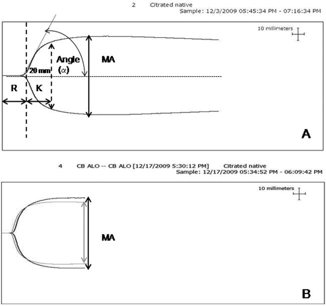
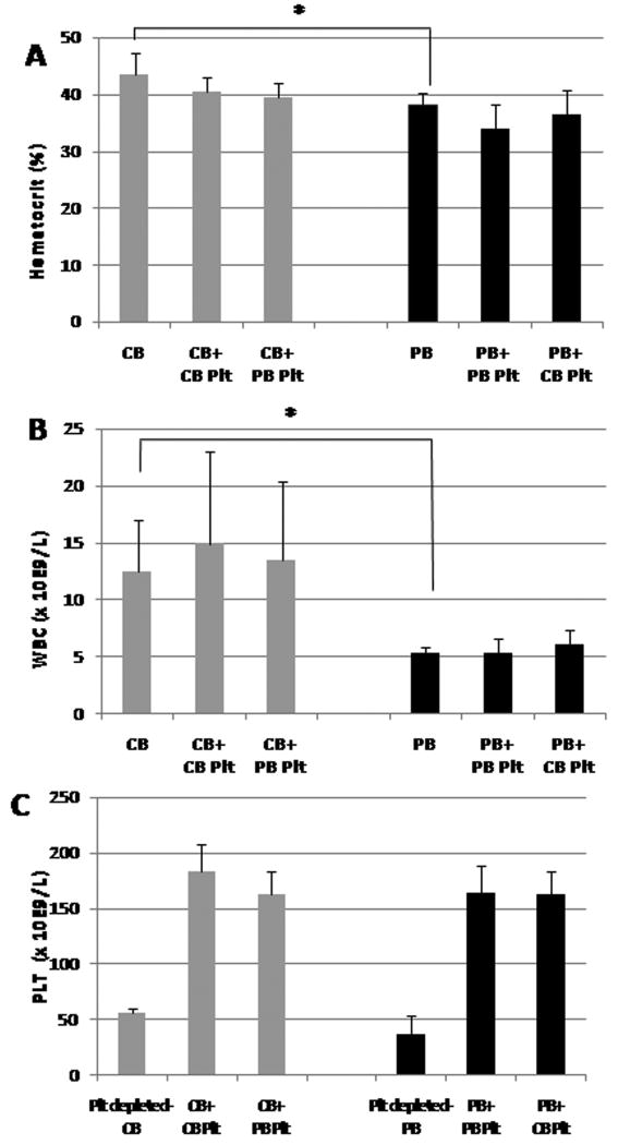
Methods: Cord blood (CB) and adult peripheral blood (PB) were separated (with a modified buffy coat method) to generate miniaturized platelet concentrates (PCs) and thrombocytopenic blood. PB-derived and CB-derived PCs (n = 7 per group) were then 'transfused'in vitro into thrombocytopenic CB and PB. The effects of autologous vs. allogeneic (developmentally mismatched) 'transfusions' were evaluated with whole blood aggregometry, a platelet function analyzer (PFA-100), and thromboelastography (TEG).

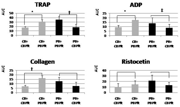
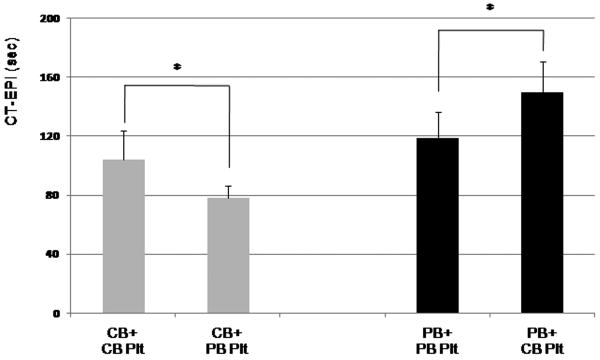
Results: Adult platelets aggregated significantly better than neonatal platelets in response to thrombin receptor-activating peptide, ADP, and collagen, regardless of the blood into which they were transfused. The 'transfusion' of adult platelets into thrombocytopenic CB resulted in shorter CTs-EPI (PFA-100) and higher clot strength and firmness (TEG) than 'transfusion' of neonatal autologous platelets.

Conclusions: In vitro'transfusion' of adult platelets into neonatal blood results in shorter CTs than 'transfusion' with neonatal platelets. Our findings should raise awareness of the differences between the neonatal and adult hemostatic system and the potential 'developmental mismatch' associated with platelet transfusions for neonatal hemostasis.


Conflict of interest statement

Disclosure of Conflict of Interests: The authors declare that they have no conflicts of interest.

Figures

Figure 1
Figure 1
In vitro’ transfusion model. Whole blood obtained from adult subjects (peripheral blood [PB]) or cord blood (CB) was separated into platelet-poor plasma (PPP), red blood cells (RBCs), and buffy coat (BC). The BC was allowed to rest and, after a soft spin, a platelet concentrate was generated from the supernatant. The pellet resulting from this spin was mixed with the PPP and RBCs from the first centrifugation to reconstitute platelet-depleted blood. PB-derived and CB-derived platelet concentrates were mixed with platelet-depleted CB or PB to mimic autologous and allogeneic transfusions (n = 7 per group). Plt, platelet; WBC, white blood cell; RC, red cells.
Figure 2
Figure 2
Thromboelastogram profile parameters and representative profiles of post-transfusion CB samples. (A) Thromboelastogram profile. Reaction time (R) is the time from the start of tracing until the curve is 2 mm wide, and represents the time to start of clot formation. Clot formation time (K) is the time measured from R to the point where the amplitude reaches 20 mm. Maximum amplitude (MA) is the widest part of the graph, and reflects clot strength; Angle (α) is the size (in degrees) of the angle formed by a line tangent to the thromboelastogram tracing, and reflects the speed of clot formation. (B) Overlapping thromboelastogram graphs showing the results of a cord blood (CB) sample transfused with neonatal platelets (gray line) vs. adult platelets (black line). Transfusion with neonatal platelets resulted in a shorter R and a lower MA
Figure 3
Figure 3
Hematocrit, white blood cell (WBC) counts and platelet concentrations in cord blood (CB) (gray bars) and peripheral blood (PB) (black bars). (A) Unmanipulated CB samples had significantly higher hematocrits than unmanipulated PB samples (*P < 0.05). Post-transfusion samples had similar hematocrits as their original whole blood sources. (B) Similarly, unmanipulated CB samples had significantly higher WBC counts than unmanipulated PB samples. Post-transfusion samples had similar WBC counts as their original whole blood sources. (C) Platelet counts in platelet-depleted whole blood and in post-transfusion CB (gray bars) and PB (black bars) samples. Plt, platelets
Figure 4
Figure 4
Impedance platelet aggregometry results obtained with the Multiplate system, following stimulation with thrombin receptor-activating peptide (TRAP), ADP, collagen, and ristocetin. Gray bars represent non-manipulated cord blood (CB) samples and black bars represent non-manipulated adult peripheral blood (PB) samples (mean ± standard deviation of seven independent samples per group). AUC, area under the curve. *P < 0.001; †P < 0.01.
Figure 5
Figure 5
Mean platelet aggregation (± standard deviation) results from thrombocytopenic cord blood (CB) samples (gray bars) and adult peripheral blood (PB) samples (black bars) transfused in vitro with neonatal (CB) or adult (PB) platelets. Adult platelets aggregated better than neonatal platelets, regardless of the blood into which they were transfused, in response to thrombin receptor-activating peptide (TRAP), ADP, and collagen. The same trend was observed in response to ristocetin, but the differences were not significant. *P < 0.001, †P < 0.01, ‡P < 0.05. AUC, area under the curve; Plt, platelets.
Figure 6
Figure 6
PFA-100 closure times following stimulation with collagen and epinephrine (CTs-EPI). Cord blood (CB) samples (gray bars) generated shorter CTs-EPI than peripheral blood (PB) samples (black bars). Within each whole blood source, transfusion with adult platelets resulted in significantly shorter CTs-EPI than with neonatal platelets. Plt, platelets. *P < 0.05.

References

    1. Castle V, Andrew M, Kelton J, Giron D, Johnston M, Carter C. Frequency and mechanism of neonatal thrombocytopenia. J Pediatr. 1986;108:749–55. - PubMed
    1. Christensen RD, Henry E, Wiedmeier SE, Stoddard RA, Sola-Visner MC, Lambert DK, Kiehn TI, Ainsworth S. Thrombocytopenia among extremely low birth weight neonates: data from a multihospital healthcare system. J Perinatol. 2006;26:348–53. - PubMed
    1. Del Vecchio A, Sola MC, Theriaque DW, Hutson AD, Kao KJ, Wright D, Garcia MG, Pollock BH, Christensen RD. Platelet transfusions in the neonatal intensive care unit:factors predicting which patients will require multiple transfusions. Transfusion. 2001;41:803–8. - PubMed
    1. Dohner ML, Wiedmeier SE, Stoddard RA, Null D, Jr, Lambert DK, Burnett J, Baer VL, Hunt JC, Henry E, Christensen RD. Very high users of platelet transfusions in the neonatal intensive care unit. Transfusion. 2009;49:869–72. - PubMed
    1. Josephson CD, Su LL, Christensen RD, Hillyer CD, Castillejo MI, Emory MR, Lin Y, Hume H, Easley K, Poterjoy B, Sola-Visner M. Platelet transfusion practices among neonatologists in the United States and Canada: results of a survey. Pediatrics. 2009;123:278–85. - PubMed

Publication types