. 2011 Apr 15;589(Pt 8):2007-26.
doi: 10.1113/jphysiol.2010.202044. Epub 2011 Feb 14.

Preferential skeletal muscle myosin loss in response to mechanical silencing in a novel rat intensive care unit model: underlying mechanisms

Affiliations

Preferential skeletal muscle myosin loss in response to mechanical silencing in a novel rat intensive care unit model: underlying mechanisms

Julien Ochala et al. J Physiol. .

Abstract

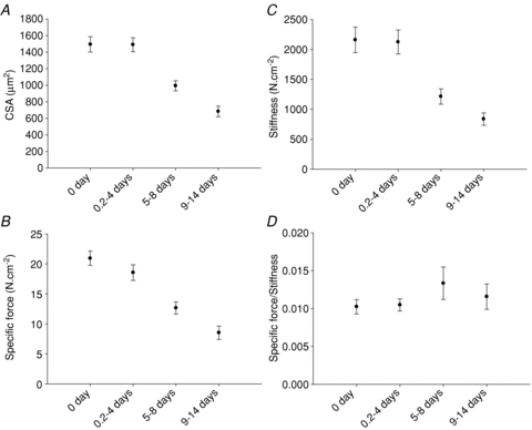
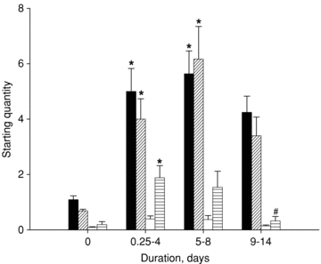
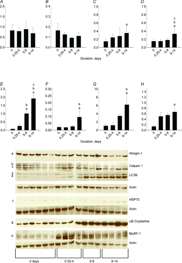
The muscle wasting and impaired muscle function in critically ill intensive care unit (ICU) patients delay recovery from the primary disease, and have debilitating consequences that can persist for years after hospital discharge. It is likely that, in addition to pernicious effects of the primary disease, the basic life support procedures of long-term ICU treatment contribute directly to the progressive impairment of muscle function. This study aims at improving our understanding of the mechanisms underlying muscle wasting in ICU patients by using a unique experimental rat ICU model where animals are mechanically ventilated, sedated and pharmacologically paralysed for duration varying between 6 h and 14 days. Results show that the ICU intervention induces a phenotype resembling the severe muscle wasting and paralysis associated with the acute quadriplegic myopathy (AQM) observed in ICU patients, i.e. a preferential loss of myosin, transcriptional down-regulation of myosin synthesis, muscle atrophy and a dramatic decrease in muscle fibre force generation capacity. Detailed analyses of protein degradation pathways show that the ubiquitin proteasome pathway is highly involved in this process. A sequential change in localisation of muscle-specific RING finger proteins 1/2 (MuRF1/2) observed during the experimental period is suggested to play an instrumental role in both transcriptional regulation and protein degradation. We propose that, for those critically ill patients who develop AQM, complete mechanical silencing, due to pharmacological paralysis or sedation, is a critical factor underlying the preferential loss of the molecular motor protein myosin that leads to impaired muscle function or persisting paralysis.


Figures

Figure 1
Figure 1. Relative change in body weight (%) in response to muscle unloading, mechanical ventilation and post-synaptic block of neuromuscular transmission
The body weight of the animals at the start of the experiment is shown in the inset.
Figure 2
Figure 2. Single muscle fibre cross-sectional area
Single muscle fibre cross-sectional area measured at a fixed sarcomere length (A), specific force (B), stiffness (C) and specific force/stiffness ratio (D) in control animals and animals exposed to NMB, muscle unloading and mechanical ventilation for durations varying between 0.25–4, 5–8 and 9–14 days. Values are means ± SEM.
Figure 3
Figure 3. Maximum velocity of unloaded shortening
Maximum velocity of unloaded shortening (V0, Muscle Lengths per second, ML . s−1) in muscle fibres expressing the type I, IIx or IIb MyHC isoform in control animals and animals exposed to NMB, muscle unloading and mechanical ventilation for 0 days (controls, black), 0.25–4 days (diagonally striped), 5–8 days (white), and 9–14 days (horizontally striped). Values are means + SEM.
Figure 4
Figure 4. Ca2+ sensitivity of force and sensitivity of stiffness
Ca2+ sensitivity of force (pCa50) and Ca2+ sensitivity of stiffness in response to different durations of NMB, muscle unloading and mechanical ventilation in muscle fibres expressing the type I (open circles), IIx (grey circles) and IIb (black circles) MyHC isoforms. Values are means ± SEM.
Figure 5
Figure 5. Fractional protein synthesis rate
Fractional protein synthesis rate in the superficial (open circles) and deep (filled circles) part of the gastrocnemius muscle in response to the different of the ICU intervention.
Figure 6
Figure 6. Actin and MyHC mRNA expression
Actin (black), MyHC type IIx (diagonally striped), IIb (white) and IIa (horizontally striped) mRNA expression in the gastrocnemius muscle from control animals and animals exposed to NMB, sedation, muscle unloading and mechanical ventilation for 0.25–4, 5–8 and 9–14 days. Values are means + SEM. **P < 0.001 (significant difference compared with controls), ##P < 0.001 (significant difference compared with 0.25–4 days group).
Figure 7
Figure 7. Myosin and actin contents
Myosin (filled) and actin (white) contents normalized to total protein content in the EDL (A), and the soleus (B). Myosin:actin ratios (C) in soleus (open circles) and EDL (filled circles) muscle cross-sections in control and mechanically ventilated animals. Values are means + SEM.
Figure 8
Figure 8. MAFbx/atrogin-1, MuRF1, calpain-1 and LC3b mRNA expression
MAFbx/atrogin-1 (black), MuRF1 (diagonally striped), calpain-1 (white) and LC3b (horizontally striped) mRNA expression in the gastrocnemius muscle from control animals and animals exposed to NMB, sedation, muscle unloading and mechanical ventilation for 0.25–4, 5–8 and 9–14 days. Values are means + SEM. *P < 0.05 (significant difference compared with controls), #P < 0.05 (significant difference compared with 0.25–4 days group).
Figure 9
Figure 9. Western blot analyses
Western blot analyses of atrogin-1 (A), 80 kDa calpain-1 (B), 75 kDa calpain (C), 18 kDa LC3 (D), 16 kDa LC3 (E), HsP70 (F), αB-crystalline (G) and MuRF1 (H) normalized to actin contents in controls (0 days) and rats exposed to post-synaptic block of neuromuscular transmission, sedation and mechanical ventilation for durations varying between 0.25–4, 5–8 and 9–14 days. Values are means + SEM.
Figure 10
Figure 10. Cross-sections of rat gastrocnemius muscle
Cross-sections of rat gastrocnemius muscle stained for the MuRF1 (red) or MuRF2 (green) and DAPI (blue). In control muscles, both MuRF1 and -2 are localised to the cytoplasm, and nuclear localisation is not prominent (arrowheads). ICU conditions induce strong nuclear translocation of both MuRFs that persists up to 9 days. At this stage, MuRFs are colocalising in the nucleus (arrowheads) but also accumulate in a perinuclear area in the cytoplasm (arrows). Scale bars, 10 μm. The overlay image for each row is shown at the left. All sections stained simultaneously and images recorded with identical gain settings. See Methods for details.
Figure 11
Figure 11. Cross-sections of rat gastrocnemius muscle
Cross-sections of rat gastrocnemius muscle stained for the MuRF2 (red), p62/SQSTM1 (green), SRF (blue) and DAPI (not shown in overlay image at left). In control muscles (A), both MuRF2 and p62 are localised to the cytoplasm, while SRF is nuclear (arrowheads). B, nuclear localisation of MuRF2 and p62 is prominent after 4 days of ICU conditions (arrowheads), where both proteins now colocalise with SRF. C, after 14 days, both MuRF2 and p62 are found in the cytoplasm with perinuclear accumulation (arrows), where they colocalise with SRF. The nuclear SRF pool is strongly depleted (arrowheads). Note the increased p62 stain, and the marked cytoplasmic signal for SRF (asterisk) at 14 days. Scale bars, 10 μm. All sections stained simultaneously and images recorded with identical gain settings. See Methods for details.

References

    1. Ahlbeck K, Fredriksson K, Rooyackers O, Maback G, Remahl S, Ansved T, et al. Signs of critical illness polyneuropathy and myopathy can be seen early in the ICU course. Acta Anaesthesiol Scand. 2009;53:717–723. - PubMed
    1. Balagopal P, Ford GC, Ebenstein DB, Nadeau DA, Nair KS. Mass spectrometric methods for determination of [13C]leucine enrichment in human muscle protein. Anal Biochem. 1996;239:77–85. - PubMed
    1. Banduseela VC, Ochala J, Chen YW, Goransson H, Norman H, Radell P, et al. Gene expression and muscle fiber function in a porcine ICU model. Physiol Genomics. 2009;39:141–159. - PubMed
    1. Bodine SC, Latres E, Baumhueter S, Lai VK, Nunez L, Clarke BA, et al. Identification of ubiquitin ligases required for skeletal muscle atrophy. Science. 2001;294:1704–1708. - PubMed
    1. Cheung AM, Tansey CM, Tomlinson G, Diaz-Granados N, Matte A, Barr A, et al. Two year outcomes, health care use and costs in survivors of acute respiratory distress syndrome. Am J Respir Crit Care Med. 2006;174:538–544. - PubMed

Publication types

MeSH terms