. 2011 Feb 21;192(4):569-82.
doi: 10.1083/jcb.201005136. Epub 2011 Feb 14.

Xenopus HJURP and condensin II are required for CENP-A assembly

Affiliations

Xenopus HJURP and condensin II are required for CENP-A assembly

Rafael Bernad et al. J Cell Biol. .

Erratum in

  • J Cell Biol. 2011 Feb 21;192(4):preceding 569
  • J Cell Biol. 2011 Mar 7;192(5):899

Abstract

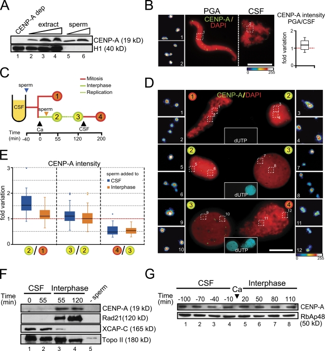
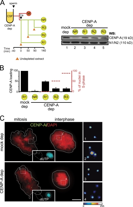
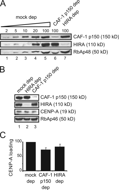
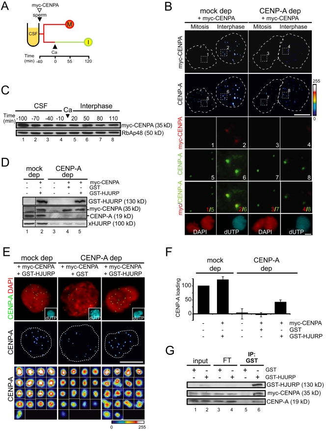
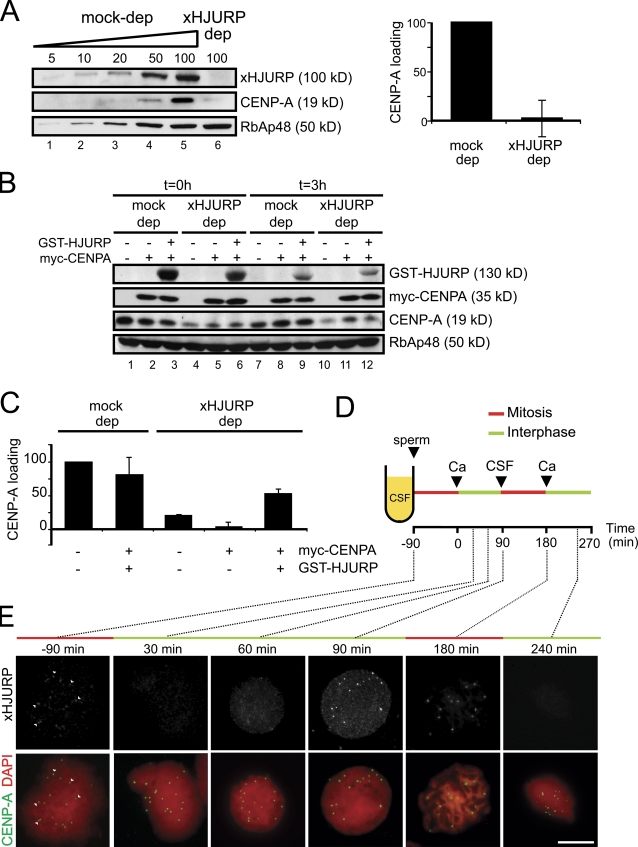
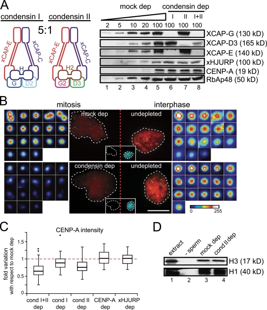
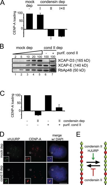
Centromeric protein A (CENP-A) is the epigenetic mark of centromeres. CENP-A replenishment is necessary in each cell cycle to compensate for the dilution associated to DNA replication, but how this is achieved mechanistically is largely unknown. We have developed an assay using Xenopus egg extracts that can recapitulate the spatial and temporal specificity of CENP-A deposition observed in human cells, providing us with a robust in vitro system amenable to molecular dissection. Here we show that this deposition depends on Xenopus Holliday junction-recognizing protein (xHJURP), a member of the HJURP/Scm3 family recently identified in yeast and human cells, further supporting the essential role of these chaperones in CENP-A loading. Despite little sequence homology, human HJURP can substitute for xHJURP. We also report that condensin II, but not condensin I, is required for CENP-A assembly and contributes to retention of centromeric CENP-A nucleosomes both in mitosis and interphase. We propose that the chromatin structure imposed by condensin II at centromeres enables CENP-A incorporation initiated by xHJURP.


Figures

Figure 1.
Figure 1.
CENP-A deposition occurs in early interphase in Xenopus egg extracts. (A) Sperm nuclei contain CENP-A. Immunoblot analysis of increasing amounts of egg extract (1, 2, and 3 µl; lanes 2–4), 3 × 10⁵ and 6 × 10⁵ sperm nuclei (lanes 5 and 6), and 1 µl of an extract depleted of CENP-A as control (lane 1). Unlike CENP-A, histone H1 is present in the soluble extract but not in the sperm nuclei. (B) Sperm nuclei were incubated with a buffer containing polyglutamic acid (PGA) or in CSF extract, mixed, centrifuged on the same coverslip, stained with anti-CENP-A (green) and DAPI (red), and imaged together. One representative pair of nuclei is shown. Bar, 10 µm. In the blown-up images on the left the intensity of the CENP-A labeling has been coded with a color gradient going from blue (minimum) to white (maximum). The intensity of CENP-A signals was measured for 15 pairs of nuclei and plotted as fold variation of the average signal for each pair. (C) Outline of the chromatin assembly experiment and the time points at which samples were taken for analysis. The time of calcium addition is considered t = 0. Black arrowheads indicate additions to the extract. The blue and orange arrowheads indicate the time of sperm addition in the two different experiments described in the main text. (D) Representative images of pairs of nuclei from two consecutive time points of the assembly reaction processed and analyzed together. Nuclei were stained with CENP-A (green) and DAPI (red). Replication was visualized by incorporation of biotin-dUTP (cyan, insets in the center). Bar, 10 µm. (E) Graph showing the fold variation of CENP-A signal intensities between two given time points (indicated by the numbers as in panel C) for two different time course experiments: blue boxes for the experiment in which sperm is added to CSF extract and orange boxes for addition of sperm in interphase extract. Data for each time point come from 15 pairs of nuclei in at least two independent experiments. (F) Immunoblot analysis of chromatin fractions obtained after incubation of sperm nuclei in CSF extract (lanes 1 and 2) or incubated in CSF for 40 min and then driven in interphase by addition of calcium (lanes 3 and 4). The times indicated correspond to the scheme in panel C. A mock assembly reaction without sperm DNA is shown as control (lane 5). (G) Immunoblot analysis of samples taken from an egg extract at the indicated times. The time of calcium addition is considered t = 0. The histone chaperone RbAp48 is shown as loading control.
Figure 2.
Figure 2.
A window of opportunity for CENP-A loading in early interphase. (A) Outline of the chromatin assembly experiment (left) and immunoblot analysis of total extracts to show soluble CENP-A levels under the indicated conditions (right). The histone binding protein N1/N2 is shown as loading control. Sperm was added at t = 0 to CSF extracts (mock depleted or CENP-A depleted). For the mock-depleted extract, one-half volume of undepleted interphase extract was added at the time of calcium addition. For the CENP-A–depleted extract, either no extract was added back (no rescue [NR] condition) or one-half volume of undepleted interphase extract was added at the times indicated (R1, R2, and R3). (B) Bar graph showing the CENP-A loading efficiency under the conditions explained above, with respect to the mock-depleted extract. A small sample of the assembly mixture was taken right before addition of undepleted extract to check the percentage of nuclei showing some incorporation of biotin-dUTP (i.e., undergoing replication, red dotted line) at the time of restoring CENP-A availability, although analysis of CENP-A incorporation was performed comparing CENP-A signals from fully replicated interphase nuclei (taken at t = 140 min) and mitotic chromosomes from images such as those shown in C. At least 15 pairs of nuclei were measured for each condition. Bars represent mean ± SE. (C) Representative images of a mass of mitotic chromosomes next to a replicated interphase nuclei stained with anti-CENP-A (green) and DAPI (red) taken from the mock-depleted (top) and CENP-A–depleted (bottom) samples. Replication was visualized by incorporation of biotin-dUTP (cyan). Bar, 10 µm.
Figure 3.
Figure 3.
CENP-A incorporation in CAF-1 p150– and HIRA-depleted extracts. (A) Immunoblot analysis of increasing amounts of a mock-depleted extract (expressed as percentage of a 1.5-µl aliquot) and 1.5-µl aliquots of CAF-1 p150– and HIRA-depleted extracts with the indicated antibodies. RbAp48 levels are also shown. (B) Immunoblot analysis of aliquots from a mock-depleted extract, HIRA- and CAF-1 p150–depleted extracts to show the remaining levels of CENP-A. RbAp46 levels are also shown. (C) Bar graph showing the CENP-A loading efficiency in the depleted extracts with respect to the mock. Bars represent mean ± SE from two independent experiments.
Figure 4.
Figure 4.
Human HJURP promotes loading of Xenopus CENP-A. (A) Outline of the chromatin assembly experiment. Mitotic chromosomes (M) and interphase nuclei (I) were assembled in mock- and CENP-A–depleted extracts containing in vitro–translated (IVT) myc-CENPA. (B) Nuclei and chromosomes were stained with antibodies against myc (top) and CENP-A (second from top), both color coded with a gradient as in Fig. 1 B. Insets 1–4 show that myc signals (red) overlap with CENP-A signals (green) in interphase nuclei of mock-depleted extracts (inset 2 and 6), whereas weak myc signals in CENP-A–depleted interphase nuclei (inset 4) are mostly background. DAPI staining (red) and biotin-dUTP (cyan) are shown in the bottom row. Bar, 10 µm. (C) IVT myc-CENP-A was added to a CENP-A–depleted extract along with sperm DNA and samples of the reaction were taken at the indicated times and analyzed by immunoblot with anti-myc and anti-RbAp48 as loading control. The time of calcium addition is considered t = 0. Cell cycle progression was monitored by chromosome morphology after DAPI staining (not depicted). (D) Immunoblot analysis of the extracts used for chromatin assembly in panel E. Antibodies against human and Xenopus HJURP were used to detect GST-HJURP (top) and endogenous xHJURP (bottom), respectively, whereas anti-CENPA was used to simultaneously detect myc-CENPA and endogenous CENP-A (middle). (E) Representative images of interphase nuclei from the indicated extracts stained with anti–CENP-A (green) and DAPI (red). Insets in the images of the first row show biotin-dUTP incorporation (cyan). In the images from the middle and bottom rows, CENP-A labeling has been color coded as in Fig. 1 B. In the bottom row, all centromeres from the nuclei presented above are shown at higher magnification. Bar, 10 µm. (F) Graph showing the loading efficiency of CENP-A in the indicated extracts compared with a mock-depleted extract. Bars represent mean ± SE from two independent experiments. (G) GST-HJURP interacts with myc-CENPA and endogenous CENP-A. Analysis of an immunoprecipitation reaction with anti-GST from an egg extract containing myc-CENPA and either GST alone or GST-HJURP. Aliquots (1.5%) from input and flow-through (FT) fractions were also analyzed.
Figure 5.
Figure 5.
xHJURP is required for CENP-A deposition. (A) Left, immunoblot analysis of increasing amounts of a mock-depleted extract (5–100% of a 1.5-µl aliquot, lanes 1–5) and a 1.5-µl aliquot of an xHJURP-depleted extract (lane 6). Right, graph showing the loading efficiency of CENP-A in an extract depleted of xHJURP. Data come from two different experiments. Bars represent mean ± SE. (B) Immunoblot analysis of mock-depleted and xHJURP-depleted extracts replenished or not with myc-CENP-A and GST-HJURP at the time of sperm addition (t = 0, lanes 1–6) and at the end of the experiment (t = 3 h, lanes 7–12). (C) Loading efficiency of CENP-A in the indicated extracts. Data come from two independent experiments. Bars represent mean ± SE. (D) Outline of the chromatin assembly reaction used to assess the localization of xHJURP throughout the cell cycle reproduced in the extract by successive additions of calcium and CSF extract. Sperm chromatin (800/µl) was added at time 0. The times at which aliquots were taken for analysis are indicated. (E) Immunofluorescent staining with anti-xHJURP (green), CENP-A (red), and DAPI (blue). White arrowheads point to HJURP signals that colocalize with CENP-A signals on CSF assembled chromosomes. Bar, 10 µm.
Figure 6.
Figure 6.
Condensin II prevents eviction of CENP-A nucleosomes from centromeres. (A) Left, subunit composition of the two condensin complexes. Right, immunoblot analysis of increasing amounts of a mock-depleted extract (expressed as percentage of a 1.5-µl aliquot) and extracts depleted of condensin I, condensin II, or both with the indicated antibodies. RbAp48 is shown as loading control. (B) Sperm chromatin was added to mock-depleted (top) and condensin-depleted (bottom) CSF extracts and incubated for 90 min. To serve as reference, sperm chromatin was added to an undepleted CSF extract, incubated for 40 min, and then calcium was added and incubation continued for 90 min. The latter samples (replicated interphase nuclei, as evidenced by the incorporation of biotin dUTP) were mixed with the former ones (mitotic chromosomes), centrifuged over coverslips, and stained with anti-CENP-A (green), DAPI (red), and streptavidin (cyan). Representative images of the photographed pairs are shown, with corresponding blown-up images of each individual centromere. (C) CENP-A signals in chromosomes assembled in depleted CSF extracts were measured in comparison with a reference sample, as described in B. The resulting numbers are expressed and plotted as fold variation relative to the average value obtained for the mock-depleted extract, arbitrarily set to 1 (red dotted line). Data from at least two independent experiments are shown for the depletion of condensins and from a single experiment in the case of CENP-A and xHJURP depletions. (D) Immunoblot analysis of chromatin fractions from assembly reactions in mock- and condensin II–depleted extracts. An aliquot of extract (lane 1) and a chromatin fraction from a mock assembly reaction with no sperm (lane 2) were also analyzed.
Figure 7.
Figure 7.
Condensin II is required for efficient loading of CENP-A. (A) Bar graph showing the CENP-A loading efficiency in the indicated extracts in two independent experiments. Bars represent mean ± SE. (B) Immunoblot analysis of increasing amounts of a mock-depleted extract (expressed as percentage of a 1.5-µl aliquot, lanes 1–5) and an extract depleted with anti-XCAP-D3 without (lane 6) or with 1:10 volume of affinity-purified condensin II (lane 7). (C) Bar graph showing the CENP-A loading efficiency in mock- and condensin II–depleted extracts with and without adding back purified condensin II. (D) Immunofluorescent staining of mitotic chromosomes assembled in mock- and condensin II–depleted extracts with antibodies against xHJURP (green) and CENP-A (red). DNA was counterstained with DAPI (blue). Bar, 10 µm. (E) Model to explain the role of xHJURP and condensin II in CENP-A dynamics. CENP-A and H3 nucleosomes are shown in green and red, respectively. Net incorporation of CENP-A will depend on the balance between addition of new CENP-A nucleosomes, a process in which xHJURP has a key role, and eviction of these nucleosomes. Condensin II prevents eviction of CENP-A nucleosomes but it is also required for their incorporation.

References

    1. Ahmad K., Henikoff S. 2001. Centromeres are specialized replication domains in heterochromatin. J. Cell Biol. 153:101–110 10.1083/jcb.153.1.101 - DOI - PMC - PubMed
    1. Allshire R.C., Karpen G.H. 2008. Epigenetic regulation of centromeric chromatin: old dogs, new tricks? Nat. Rev. Genet. 9:923–937 10.1038/nrg2466 - DOI - PMC - PubMed
    1. Bernad R., Sánchez P., Losada A. 2009. Epigenetic specification of centromeres by CENP-A. Exp. Cell Res. 315:3233–3241 10.1016/j.yexcr.2009.07.023 - DOI - PubMed
    1. Bloom K. 2007. Centromere dynamics. Curr. Opin. Genet. Dev. 17:151–156 10.1016/j.gde.2007.02.009 - DOI - PubMed
    1. Blower M.D., Sullivan B.A., Karpen G.H. 2002. Conserved organization of centromeric chromatin in flies and humans. Dev. Cell. 2:319–330 10.1016/S1534-5807(02)00135-1 - DOI - PMC - PubMed

Publication types

MeSH terms