. 2011 Apr 1;286(13):11155-62.
doi: 10.1074/jbc.M110.217349. Epub 2011 Feb 14.

Chronic inhibition of pyruvate dehydrogenase in heart triggers an adaptive metabolic response

Affiliations

Chronic inhibition of pyruvate dehydrogenase in heart triggers an adaptive metabolic response

Kari T Chambers et al. J Biol Chem. .

Abstract

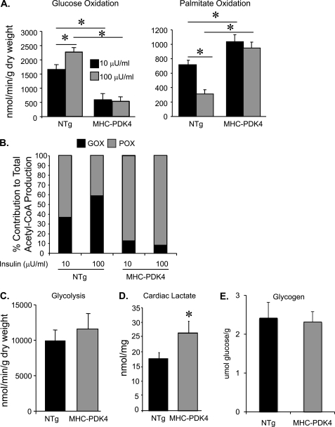
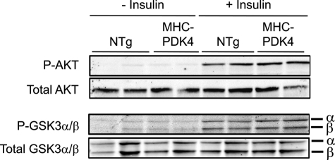
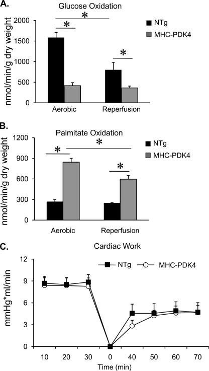
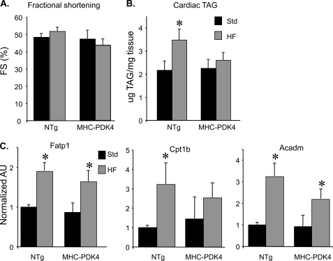
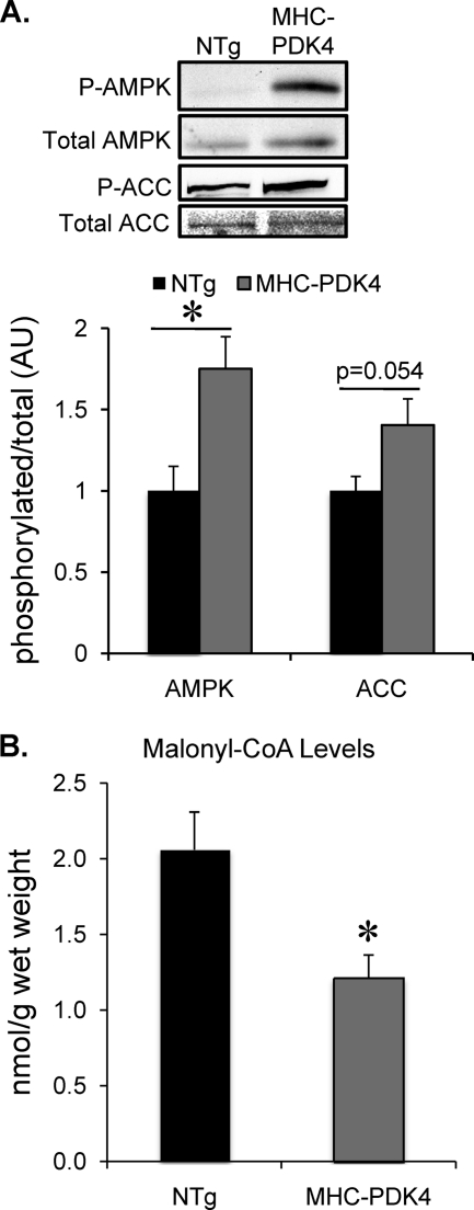
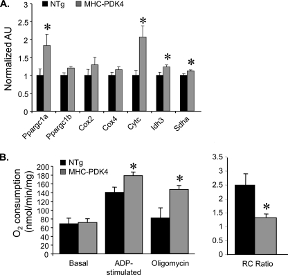
Diabetic cardiac dysfunction is associated with decreased rates of myocardial glucose oxidation (GO) and increased fatty acid oxidation (FAO), a fuel shift that has been shown to sensitize the heart to ischemic insult and ventricular dysfunction. We sought to evaluate the metabolic and functional consequences of chronic suppression of GO in heart as modeled by transgenic mice with cardiac-specific overexpression of pyruvate dehydrogenase kinase 4 (myosin heavy chain (MHC)-PDK4 mice), an inhibitor of pyruvate dehydrogenase. Hearts of MHC-PDK4 mice were shown to exhibit an insulin-resistant substrate utilization profile, characterized by low GO rates and high FAO flux. Surprisingly, MHC-PDK4 mice were not sensitized to cardiac ischemia-reperfusion injury despite a fuel utilization pattern that phenocopied the diabetic heart. In addition, MHC-PDK4 mice were protected against high fat diet-induced myocyte lipid accumulation, likely related to increased capacity for FAO. The high rates of mitochondrial FAO in the MHC-PDK4 heart were related to heightened activity of the AMP-activated protein kinase, reduced levels of malonyl-CoA, and increased capacity for mitochondrial uncoupled respiration. The expression of the known AMP-activated protein kinase target, peroxisome proliferator-activated receptor γ coactivator-1α (PGC-1α), a master regulator of mitochondrial function and biogenesis, was also activated in the MHC-PDK4 heart. These results demonstrate that chronic activation of PDK4 triggers transcriptional and post-transcriptional mechanisms that re-program the heart for chronic high rates of FAO without the expected deleterious functional or metabolic consequences.


Figures

FIGURE 1.
FIGURE 1.
Hearts of MHC-PDK4 mice exhibit increased rates of palmitate oxidation and concomitant decreased rates of glucose oxidation. A, substrate utilization rates in working hearts isolated from female NTg and MHC-PDK4 hearts (n = 5–6 per group) is shown. The bars denote mean (±S.E.) glucose oxidation (left) and palmitate oxidation (right) rates under low insulin (10 μm, black bars) or high insulin (100 μm, gray bars) perfusion conditions. B, the bars denote percent contribution to total acetyl-CoA production by glucose oxidation (GOX, black portion) or palmitate oxidation (POX, gray portion) (n = 5–6 per group). C, the bars denote mean rates of glycolysis in NTg and MHC-PDK4 hearts (n = 8–9 per group). D, bars denote mean levels of cardiac lactate in NTg and MHC-PDK4 hearts (n = 9–11 per group). E, bars denote mean levels of glycogen in NTg and MHC-PDK4 hearts (n = 8–10 per group). *, p < 0.05.
FIGURE 2.
FIGURE 2.
Insulin receptor-mediated signaling is unchanged in MHC-PDK4 hearts. Male NTg and MHC-PDK4 mice were injected IV with (10 milliunits/g of body weight) insulin or saline (−insulin), and hearts were harvested 5 min later (n = 6 per group). Representative Western blots using heart lysates are shown.
FIGURE 3.
FIGURE 3.
MHC-PDK4 hearts have normal functional recovery after ischemia-reperfusion injury. Female NTg and MHC-PDK4 hearts were subjected to ischemia-reperfusion in the isolated working preparation as described under “Experimental Procedures” with 5 mm glucose, 0.4 mm palmitate, and 100 microunits/ml insulin. A, bars represent mean glucose oxidation rates (±S.E.) under aerobic (n = 11–15 per group) conditions and after reperfusion (n = 6–12 per group). B, bars denote palmitate oxidation rates (± S.E.) under aerobic (n = 11–15 per group) conditions and after reperfusion (n = 6–12 per group). C, the line graph depicts cardiac work (±S.E.) over time during the ischemia-reperfusion protocol in NTg (black squares) and MHC-PDK4 (open circles) hearts. Time 0 represents the beginning of the reperfusion period. *, p < 0.05.
FIGURE 4.
FIGURE 4.
MHC-PDK4 mice are protected from HF diet-induced TG accumulation in the myocardium. A, echocardiography was performed on male NTg and MHC-PDK4 mice (n = 4–6 per group after standard or high fat chow diet). Bars represent mean percent left ventricular fractional shortening (FS) % (±S.E.) after 4 weeks of HF diet (gray bars) versus standard chow (black bars). B, bars denote mean cardiac triglyceride (TG) levels (± S.E.) in male NTg and MHC-PDK4 hearts after 4 weeks of either standard (black bars) or HF chow (gray bars) (n = 6–9 per group). C, levels of mRNAs as determined by quantitative real-time-PCR were performed on total RNA isolated from heart ventricles of male mice from the genotypes indicated (n = 6–9 per group). Values are shown as arbitrary units (AU) normalized (=1.0) to the NTg on standard chow. *, p < 0.05.
FIGURE 5.
FIGURE 5.
Activation of AMPK and decreased malonyl-CoA levels in hearts of MHC-PDK4 mice. A, top, representative Western blot analyses were conducted on lysates of heart ventricles of female mice of indicated genotypes for phosphorylated (P) and total AMPK and acetyl-CoA carboxylase (ACC) as denoted. Bottom, bars represent the ratio in arbitrary units (AU) of phosphorylated/total levels of AMPK or acetyl-CoA carboxylase as indicated normalized (=1.0) to the NTg (n = 7 per group). *, p < 0.05 compared with NTg. B, bars represent the mean (±S.E.) cardiac malonyl-CoA levels as determined from heart ventricles of male mice from indicated genotypes (n = 6–9 per group). *, p < 0.05 compared with NTg.
FIGURE 6.
FIGURE 6.
Induction of PGC-1α gene expression and increased respiratory uncoupling in mitochondria isolated from MHC-PDK4 hearts. A, levels of specific mRNAs based on quantitative real-time-PCR performed on RNA isolated from heart ventricles of male mice from the genotypes indicated (n = 8–10 per group) are shown. Bars represent arbitrary uints (AU) normalized (=1.0) to the NTg. B, mitochondrial respiration rates determined by oxygen consumption (VO2) performed on mitochondria isolated from ventricle of male NTg and MHC-PDK4 hearts are shown. Mean values (±S.E.) are shown for the basal, ADP-stimulated, and oligomycin-inhibited states using succinate/rotenone as a substrate. The mean respiratory control (RC) ratio (±S.E.) is shown on the right. *, p < 0.05 compared with NTg.

References

    1. Engelgau M. M., Geiss L. S., Saaddine J. B., Boyle J. P., Benjamin S. M., Gregg E. W., Tierney E. F., Rios-Burrows N., Mokdad A. H., Ford E. S., Imperatore G., Narayan K. M. (2004) Ann. Intern. Med. 140, 945–950 - PubMed
    1. Zimmet P., Alberti K. G., Shaw J. (2001) Nature 414, 782–787 - PubMed
    1. Fein F. S., Sonnenblick E. H. (1985) Prog. Cardiovasc. Dis. 27, 255–270 - PubMed
    1. Regan T. J., Lyons M. M., Ahmed S. S., Levinson G. E., Oldewurtel H. A., Ahmad M. R., Haider B. (1977) J. Clin. Invest. 60, 884–899 - PMC - PubMed
    1. Rubler S., Dlugash J., Yuceoglu Y. Z., Kumral T., Branwood A. W., Grishman A. (1972) Am. J. Cardiol. 30, 595–602 - PubMed

Publication types

MeSH terms

Substances